Small Pars Intermedia Cyst
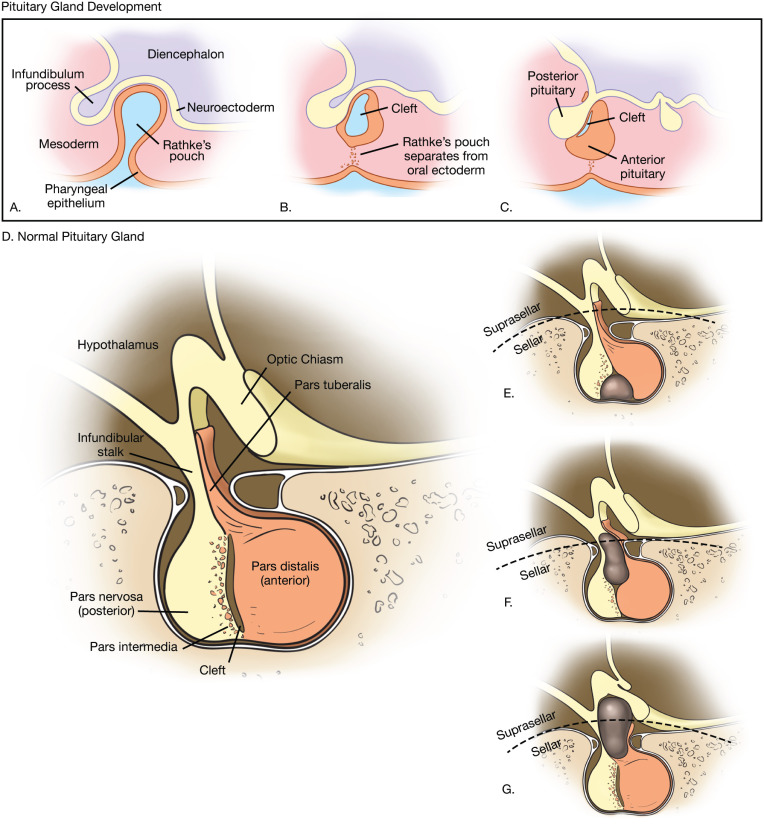
The pars intermedia refers to the cleft that normally develops separating the anterior and posterior pituitary. Rathkes cleft cysts often may be difficult to differentiate from other intrasellar or suprasellar masses on radiologic studies.
Pituitary Cysts Pituitary World News

The pars intermedia is very poorly developed in the human pituitary but is prominent in the pituitaries of most other mammals.
Small pars intermedia cyst. Pars intermedia cysts include any pituitary cyst that develops on the pars intermedia region of the pituitary gland. These cysts are usually small measuring less than 5 mm in diameter. Small pars intermedia cysts clinically silent pituitary infarcts and foci of necrosis may be incidental findings and may be confused with microadenomas by both imaging techniques. The cysts are the remainder of Rathkes pouch. This happens when the cleft opens up or maybe never even closes and a little tiny cystic structure forms. The most common cyst is a Rathkes cleft cyst RCC.
The goals in imaging patients with presumed pituitary macroadenoma include diagnosis differential diagnosis. They typically lie in the pars intermedia between the anterior and posterior pituitary lobes. Perhaps the most common abnormality of this area is what is known as a pars intermedia cyst. These usually measure less than 3 mm in size and are of no consequence. For example in the monkey pituitary you will see the pars intermedia as a prominent layer several cells thick lying between the. But a small cyst detected in this area cant easily be distinguished from.
Brain magnetic resonance imaging MRI scans reveal incidental pars intermediaRathke cleft pituitary cysts or cyst-like structures so frequently that the American College of Radiology ACR has developed management guidelines for adult patients. These colloid-filled cysts are common particularly in children in whom the area is better defined but they often cause no symptoms because they are small in size. One case of infundibular thickening in an asymptomatic adult appeared to be secondary to a vascular malformation resulting in venous congestion. The pars intermedia in a thin layer of epithelial cells located between pars distalis and neurohypophysis. INH was the most common inflammatory lesion. Pituitary pars intermediaRathke cleft cysts or cyst-like structures are commonly encountered in children undergoing brain magnetic resonance imaging MRI especially when examinations include thin-section high-resolution sequences.
A retrospective review of MR studies. A Rathke cleft cyst develops from a piece of the fetus developing Rathke pouch which ultimately becomes part of the pituitary gland. Pars intermedia is the boundary between the anterior and posterior lobes of the pituitary. Pars intermedia cysts are usually small and cause no symptoms or health problems. 177 When they enlarge they may become symptomatic causing hypopituitarism or more rarely diabetes insipidus. They develop while a fetus is growing in the womb.
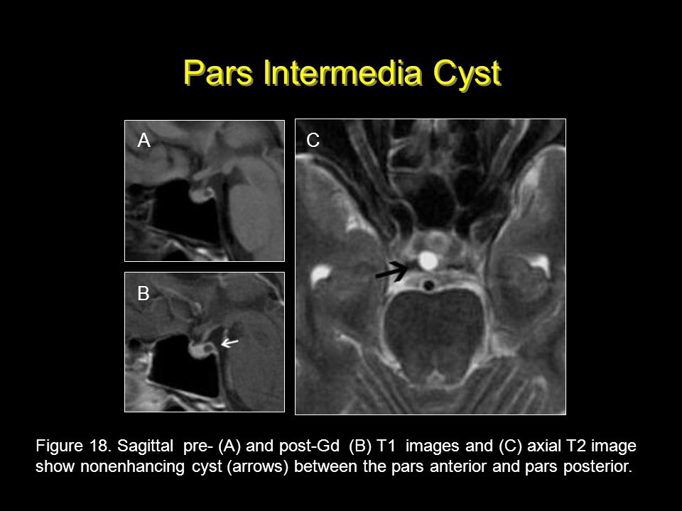
Symptomatic Rathkes cleft cysts usually present in adulthood. INH Inflammatory and infectious infundibular lesions were found exclusively in adults. In the human the intermediate lobe of the pituitary is vestigial and often includes small cystic spaces derived from the cleft. The purpose of this study was to describe the significance of intracystic nodules a diagnostic characteristic found in Rathkes cleft cysts on MR images. Postcontrast T1 sagittal MRI of 2 different patients showing small nonenhancing cyst white arrows less then 3 mm in size between the anterior and posterior pituitary gland consistent with pars intermedia cyst. Most are remnants of the craniopharyngeal Rathkes pouch and are distinct from Rathkes cleft dilation that is localized between the pars distalis and pars intermedia.
2 Pars intermedia cyst. Pituitary cysts in the pars distalis are frequent incidental findings in rats more than a year old and in mice. Rathke cleft or pars intermedia cysts. To determine the prevalence of pituitary cystic lesions in children at our institution using modern MRI technique. They are seen with regularity on pituitary MRIs if one actually looks for them. Rathke cleft cysts with or without the squamous metaplasia and the pars intermedia of the pituitary gland expressed simple epithelium cytokeratins CK7 CK8 CK19 and CK20 and most of them did not express CK18.
Incidentally detected pituitary cysts in children common on MRI. Up to 20 cash back The 3 mm pars intermedia cyst is a very small cyst and these types of cysts generally remain the same size throughout the life or sometimes even spontaneously resolve too. It contains three types of cells - basophils chromophobes and colloid -filled cysts. This is a cyst that occurs in the pituitary gland region and this is a congenital type of cyst means this is present from the birth itself. Epithelium-lined cysts are present in the pars nervosa. Symptomatic cysts can cause headaches and growth malfunctions that can mimic thyroid problems.
Rathkes cleft cysts are a type of pars intermedia cyst. An evaluation of 1000 nonselected autopsy specimens revealed that 113 of the pituitary glands 113 harbored incidental Rathke cleft cysts 2. Macroadenomas are well seen on MRI as they are on CT. It arises from the posterior wall of Rathkes pouch and contains vestigial lumina of Rathkes pouch which appear as narrow vesicles of variable length. In humans an improper degradation of the Rathkes pouch can cause a pars intermedia cyst. Others include arachnoid cysts pars intermedia cysts and epidermoid cysts.
Rathke cleft cysts are non-cancerous fluid-filled growths that develop between the parts of the pituitary gland at the base of the brain. Their similar cytokeratin expression patterns suggest that Rathke cleft cysts arise from the Rathke pouch remnants. BACKGROUND AND PURPOSE. Rathkes cleft is a normal part of the developing pituitary gland in the fetus. In some people the cleft becomes filled with liquid creating a Rathkes cleft cyst.
Congenital Abnormalities Of The Sellar And Parasellar Regions Ppt Video Online Download

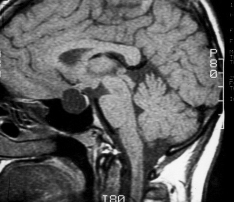
Sagittal Fspgr Bravo Image Displaying A 4 Mm Pars Intermedia Pituitary Download Scientific Diagram
Pubs Rsna Org
Pituitary Cysts In Childhood Evaluated By Mr Imaging American Journal Of Neuroradiology

Pubs Rsna Org
Rathke Cleft Cyst Radiology Key
Pubs Rsna Org

Normal Histology

Rathke S Cleft Cyst Neurosurgery
Mr Imaging Findings Of Rathke S Cleft Cysts Significance Of Intracystic Nodules American Journal Of Neuroradiology

Pituitary Cysts In Childhood Evaluated By Mr Imaging American Journal Of Neuroradiology

Figure 4 Sagittal Enhanced T1 Weighted Image Endotext Ncbi Bookshelf
Pens Org
Rathke S Cleft And Other Pituitary Cysts Pituitary World News
Posting Komentar untuk "Small Pars Intermedia Cyst"