Cystic Lesion In Inguinal Canal Radiology
Cystic or encapsulated fluid collections are relatively common benign lesions. In the reported second and third cases clinical history physical examination and ultrasonography raised the suspicion of cystic lymphangiomas.

Scielo Brasil Contents Of The Inguinal Canal Identification By Different Imaging Methods Contents Of The Inguinal Canal Identification By Different Imaging Methods
Cystic lesion in the inguinal canal with posterior acoustic attenuation.

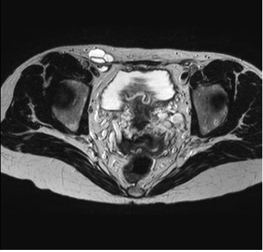
Cystic lesion in inguinal canal radiology. It showed a well-defined cystic sac-like tubular lesion in the left inguinal region extending into the left labia majora. The reported case was a cystic thin-walled structure in the inguinal area with hypointense T1 signal and hyperintense T2 signal characteristics. Medial to external iliac vessels and origins of inferior epigastric arteries. Walter and Martin described a case of a cyst of the canal of Nuck as a cyst within a cyst which is similar to their description of a spermatic cord. It was the first report of MRI findings in this pathology in English literature. They are even more frequent with an incidence of 75.
3 This case is a unique presentation of bilateral canal of Nuck cysts. UltrasonographyUS of the scrotum has been demonstrated to be useful in the diagnosis of fluid in the scrotal sac. Canal of Nuck cyst is an inguinal cyst in females. Round ligaments seen on cranial extent leading into them. Can show peripheral enhancement. This lesion measured approximately 55 cm craniocaudal 18 cm transverse 10 cm anteroposterior in dimension.
Hypodense lesion with our without internal septations. Incisive canal cysts also known as nasopalatine duct cysts NPDC are developmental non-neoplastic cysts arising from degeneration of nasopalatine ducts. Inguinal hernia containing fluid in. Magnetic resonance imaging MRI was advised for further evaluation. Ultrasonography is the primary diagnostic imaging modality. Cysts in the canal of Nuck are rare with about 400 cases reported worldwide.
At MR imaging seminal vesicle cysts are seen as well-defined intraseminal unilocular round or oval cystic lesions posterior to the urinary bladder with variable signal intensity on T1-weighted images and most often fluid signal intensity on T2-weighted images. Right cyst is elliptical while left cyst is more ovoid. According to Holden et al. Grayscale US characterizes the lesions as testicular or extratesticular and with color Doppler power Doppler and pulse Doppler any perfusion can also be assessed. Generally be categorized as mesenchymal tumors skin appendage lesions metastatic tumors other tumors and tumorlike lesions or inflammatory. The variety of cystic lesions of the inguinal and scrotal region as well as the possibility of intra-cystic hemorrhage or inflammation impede the precise pre-operative diagnosis of a cystic lymphangioma 2 3 5 13.
Right cyst has internal coarse calcification possibly from previous infectioninflammation. Scrotal and inguinal cystic lymphangiomas are treated with surgical excision with care to preserve the intra-scrotal structures and the structures of the inguinal canal. Hydrocele of the Nuck canal cyst of the Canal of Nuck or female hydrocele are equivalent terms for a rare developmental disorder of the reproductive system of women and accounts for a limited number of cases of benign painless or painful swelling in the inguinal region or even to the labia majora 4 5. Cysts of the epididymis are the most common cystic lesions and they are diagnosed in 2040 of asymptomatic patients undergoing US examination. The majority of these are unilateral at presentation and most commonly occur in young women. Bilateral cystic lesions in inguinal canals.
To review the reliability of computed tomographic CT and magnetic resonance MR imaging features in distinguishing lipoma and well-differentiated liposarcoma. Axial and coronal T2-weighted MRI sequences A and B respectively showing a lesion with a cystic aspect in the right inguinal canal with thin walls no septa and no solid components consistent with a cyst of the canal of Nuck. Superficial soft-tissue masses can. The cyst contains some thin septa hypointense on T2-weighted images that give it an aspect of crown of the rosary at the inguinal orifice Fig. May be further divided into lesions that arise in association with the. Complete excision is necessary to prevent recurrence.
T1 isointense T2 hyperintense typical appearance of a cystic lesion. They are usually not palpable at clinical examination due to their small size and softness. With regard to their imaging features these masses. This cyst contained a heterogeneous tubular structure. MRI showed a pear-shaped cyst with a superior pedicle continuing in the inguinal canal along the course of the round ligament of the uterus. These ducts usually regress in fetal life.
Differentials for Canal of Nuck cyst.
Hong Kong College Of Radiologists Answer Of September 2014

Ultrasound Image At The Level Of Right Inguinal Canal Showing Anechoic Download Scientific Diagram

Sonography Of Various Cystic Masses Of The Female Groin Oh 2007 Journal Of Ultrasound In Medicine Wiley Online Library

Sonographic Findings Of Groin Masses Yang 2007 Journal Of Ultrasound In Medicine Wiley Online Library
Onlinelibrary Wiley Com

Axial Mri Of The Right Inguinal Region The Hydrocele Of The Canal Of Nuck Download Scientific Diagram

Canal Of Nuck Cyst Hydrocele Radiology Case Radiopaedia Org

Magnetic Resonance Findings Of A Hydrocele Of The Canal Of Nuck A Download Scientific Diagram

Scielo Brasil Contents Of The Inguinal Canal Identification By Different Imaging Methods Contents Of The Inguinal Canal Identification By Different Imaging Methods
Inguinal Sonography Ultrasonography Of The Right Inguinal Area Shows A Download Scientific Diagram

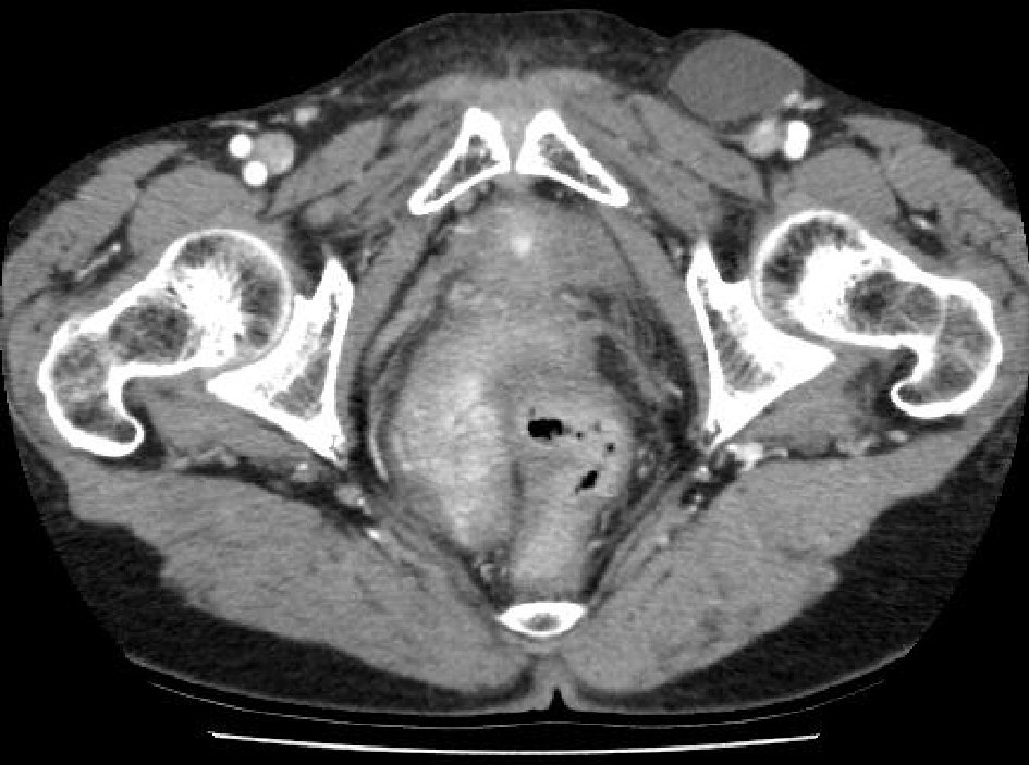
Ct Scan Demonstrating A Lobular Cystic Mass In The Inguinal Canal Download Scientific Diagram
Hong Kong College Of Radiologists Answer Of September 2014
A Mesothelial Cyst Presenting As Inguinal Mass Two Case Reports And Literature Review Choi Journal Of Current Surgery

Palpable Nonreducible Cystic Mass In The Inguinal Region Located Download Scientific Diagram
Posting Komentar untuk "Cystic Lesion In Inguinal Canal Radiology"