Carotid Artery Calcification X Ray
Ad See Doctors with Extensive Experience in Carotid Artery Numbness. The sensitivity of carotid calcification in detecting clinically significant stenosis assuming any calcification is abnormal was 89 with a specificity of 46.
Onlinelibrary Wiley Com
In these community-dwelling older adults without clinical CVD internal carotid artery intima-media thickness was most closely related to CAC.

Carotid artery calcification x ray. When calcium or calcification in the carotid arteries is seen on dental X-rays it means you have a high risk for a CV cardiovascular event like a heart attack or a stroke. 1 Carotid artery plaques which typically develop in the area of. Calcification of the Arteries There are many different ways that calcification can occur. Finding Carotid Plaque On A Standard Dental X-Ray May Predict Fatal Heart Attack Or Stroke UB Study Finds. Calcifications in the area of the carotid arteries are depicted in 3 to 15 of panoramic radiographs PRs exposed in different populations1 2 3 One study reported that 99 of carotid calcifications depicted on PRs coincide with ultrasound-verified calcified carotid plaques CCPs of various sizes. With diligent diet and supplementation you may be able to reverse the.
The carotid artery calcification score is calculated using computed tomography angiography CTA which is a confirmatory test that enables the examination of plaque composition and computation of the carotid artery calcification score. Atheroma-related formations of thrombi and emboli in the carotid artery is the most frequent cause of stroke. In one study 1 in 4 men with dental X-rays showing carotid artery calcium deposits had a CV event within 35 yrs. However 175 of those with a CAC or 400 would be missed in the ascertainment of subclinical atherosclerosis using the previously published composite o. The differential diagnosis are carotid artery atheromas calcified submandibular lymphnodes and sialoliths of the submandibular gland. Calcifications within the carotid arteries will appear lateral to the spine whereas calcifications in.
As CAC can be asymptomatic it is critical to identify it early using diagnostic imaging. Calcifications in the region of the 2nd 3rd and 4th cervical vertebrae may often appear on panoramic radiographs PRs which are a routine and indispensable part of dental examinations8 9 Several studies have evaluated the use of PRs in the detection of carotid artery calcifications CACs and the correlation between findings from PRs. The prevalence of intracranial artery calcification are. Carotid calcifications from calcified triticeous or thyroid cartilages calcified lymph nodes and non-carotid phleboliths. Ad Learn more about the signs that may reveal you have an Issue that need attention. Carotid Artery Calcification CAC The use of panoramic x-ray technology adopted by the best cosmetic dentists in the world has led to an exciting tool that helps screen for possible heart conditions.
Panoramic radiography is a basic diagnostic tool in the dental field where calcifications are seen occasionally in the lateral parts of the x-ray. Friedlander and Baker 6 noted that by panoramic radiographs can be identified asymptomatic patients at risk for stroke. The question arises whether the presence of bone and extensive sheets of calcification in the carotid artery. 8 For this reason it is important to have an Anterior-Posterior AP radiograph of the neck made using soft tissue exposure settings. Although patent aneurysms may contain mural calcification partially or completely thrombosed aneurysms commonly. Friedlander and Lande 5 were the first to describe the presence of calcifications in the region of common carotid artery by panoramic radiographs performed in the routine dental diagnosis.
Such calcification can be seen in regular oral panoramic radiography. Other causes of vascular intracranial calcifications include. Its very existence indicates that the patient has atheroma which may result in both ischemia and a cerebrovascular event. Calcification in the Carotid Artery There is no question that calcification in the carotid artery is undesirable. Our Carotid Artery Test Identifies Plaque Build Up Risk of Stroke Vascular Disease. The main cause leading to this condition is arteriosclerosis a hardening and thickening of the arterial walls often accompanied by the buildup of fatty deposits atherosclerosis and artery calcification CAC.
Learn What Sets Mayo Clinic Apart. High vegetable oil intake blood thinning medications high fluoride intake vitamin C deficiency a high calcium intake without adequate magnesium K and D and excessive vitamin D to low vitamin K intake. 21 Calcification is a complication in the evolution of atheromatous plaque enabling potential. Strokes heart attacks and the potential of brain damage are some of the health issues. Carotid artery calcification CAC identified in panoramic radiographs PRs have a prevalence. Middle cerebral artery 5.
The hyoid bone styloid process triticeal cartilages thyroid cartilage epiglottis calcifications in the stylo-hyoid and stylomandibular ligaments tonsilloliths. Ad Ultrasound Technology Detects Risk For Carotid Artery Blockage. When getting a full panoramic x-ray at the dentist arteries in the neck can show CAC buildup in the arteries before a stroke occursBy locating possible CAC. Atherosclerotic conditions of a carotid artery can lead to neural ischemia inadequate oxygen to the brain thus resulting in a cerebrovascular accident CVA or stroke. Retrieved December 26 2021 from. The good thing is that carotid artery calcification can be spotted in dental panoramic x-ray images which can be used for CAC screening.
Prior research has already shown that carotid artery calcification. The differential diagnosis of the images of carotid artery atheromas in panoramic radiography must be made considering a series of anatomic structures in the cervical and adjacent regions such as. 4007 panoramic radiographs 100 from patients 40 years were scanned retrospectively. University At Buffalo. The likelihood ratios for 50 stenosis by angiography varied from 024 no calcification to 341 level III and for 50 stenosis by duplex ultrasound varied from 021 no calcification to more than 587 level III.

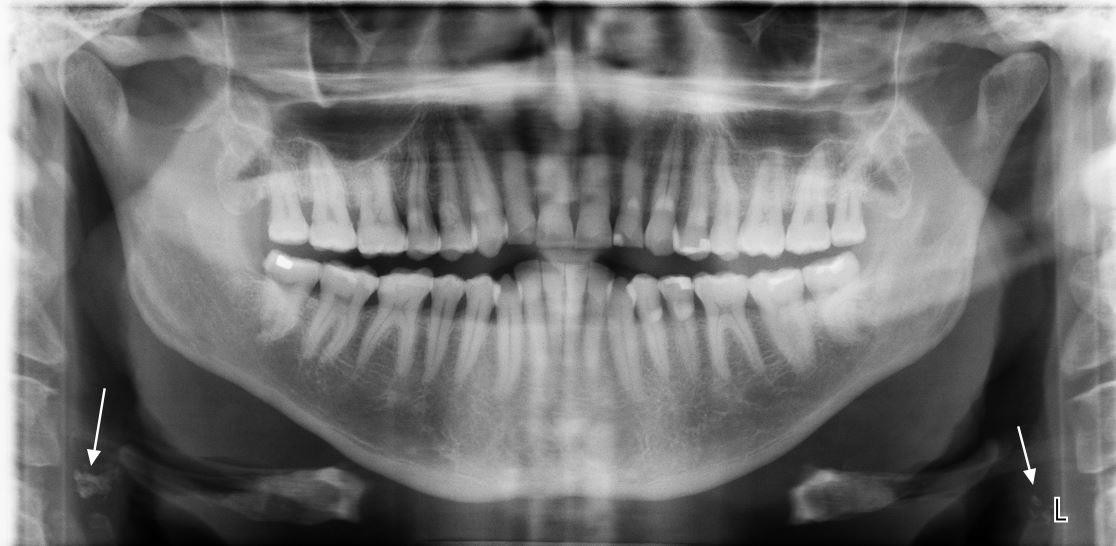
A Typical Carotid Artery Calcification Visible On The Left Side Of The Download Scientific Diagram

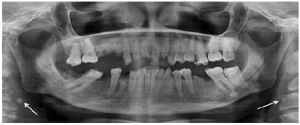
Panoramic Radiograph Of Carotid Artery Calcifications Visible On The Download Scientific Diagram
Unilateral Carotid Artery Calcification Download Scientific Diagram
Prevalence Of Carotid Artery Calcification Detected On Panoramic Radiographs Of Patients With A History Of Hypertension Or Myocardial Infarction
Diseases Free Full Text Carotid Artery Calcification A Digital Panoramic Based Study Html

Figure 2 From Radiation Associated Carotid Artery Atherosclerosis Case Report And Review Of Contemporaneous Literature Semantic Scholar
Colgate Oral Health Network Free Dental Continuing Education
Colgate Oral Health Network Free Dental Continuing Education

Beware Of Calcification In The Neck

Antero Posterior Radiograph With Arrows Pointing Out The Carotid Artery Download Scientific Diagram

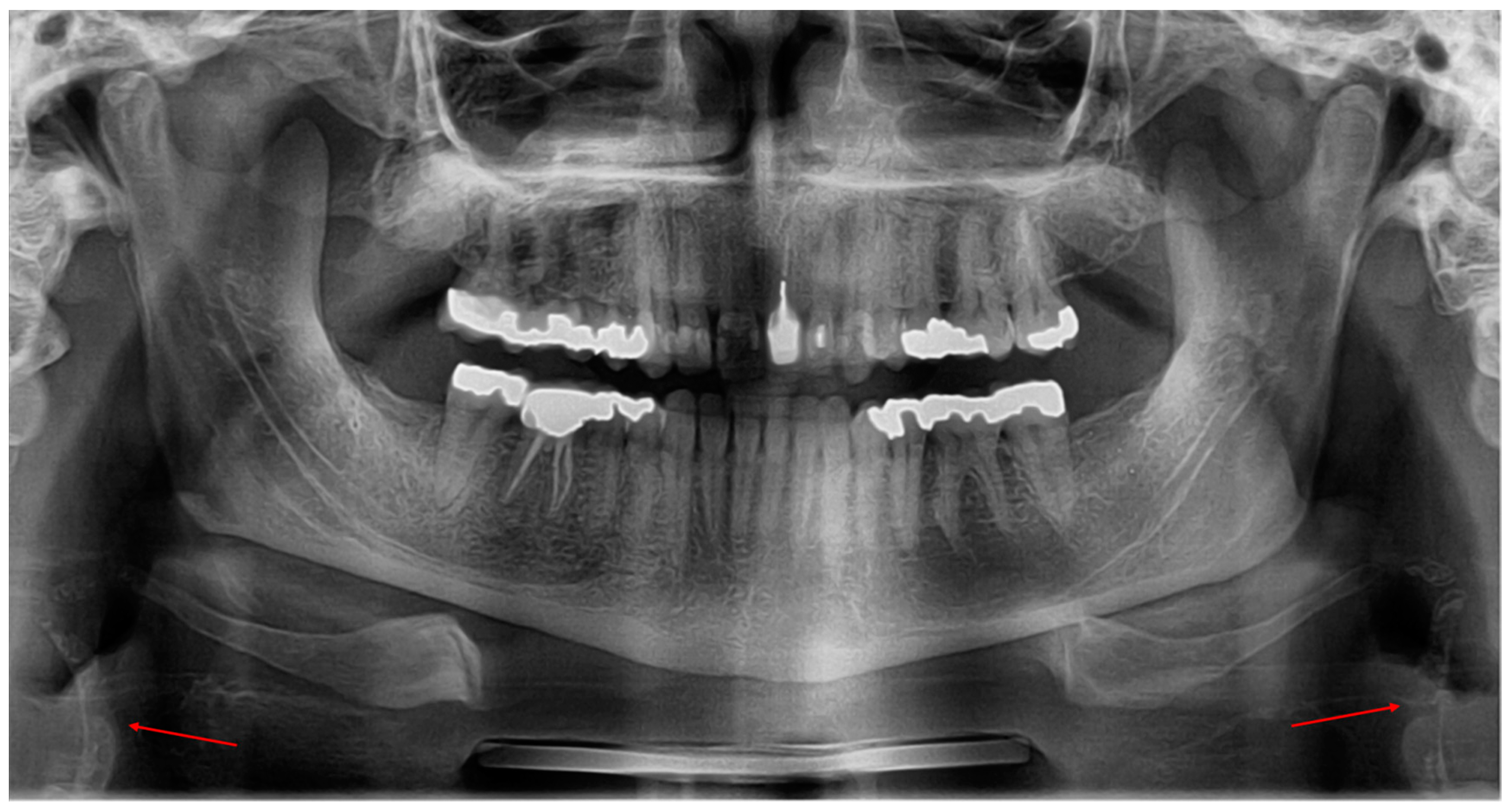
Carotid Artery Calcifications Visible On The Right And Left Neck Download Scientific Diagram

Calcified Carotid Artery Atheroma And Stroke The Journal Of The American Dental Association

Radiology Flashcards Quizlet

Carotid Calcium Cardiosound
Posting Komentar untuk "Carotid Artery Calcification X Ray"