Does Lumbar Mri Include Sacrum
And yes occasionally Ive reported incidental findings in an abdominal organ when interpreting an MRI of the LS spine. When the clinical examination and referral notes are inadequate Back Pain FI the cause of the pain may well be sacral rather than lumbar.
Lumbosacral Spine Mri Radiology Key
Indications for the procedure.

Does lumbar mri include sacrum. Cover L4-5 to tip of the coccyx through SI Joints and Sacrum. Further it is followed by the sacral section consisting of 5 joints in one common bone and the coccyx a rudimentary organ similar in structure to the sacrum but smaller in size. This means that it includes the lumbar spine and the sacrum but typically does NOT include the tailbone coccyx. However even normal appearing joints can be painful. You would need a hip specific mri. 75557 - Cardiac MRI for morphology and function wo contrast 75559 - wstress imaging 75561 - Cardiac MRI for morphology and function w and wo contrast and further seques 75563 - wstress imaging.
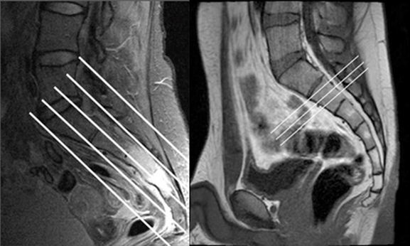
Include lateral femoral head margins. No need for entire pelvis. The lumbosacral spine is made up of the five lumbar vertebral bones L1 thru L5 the sacrum the bony shield at the bottom of your spine and the coccyx tailbone. Angle parallel to long axis of sacrum perpendicular to Axial Obl. Use of contrast on MRI may be useful to determine vascularity of tumor and whether further study is. MRI of the sacral or sacro-coccygeal spine involves the identification of pathological changes in the coccyx and in the sacrum and sacroiliac joints where there are multiple blood vessels and nerve roots that provide innervation to.
Orthopedicextremity exams on reverse. Measurements mainly focus on a change in signal intensities and less on absolute distances or angles. 4037774MRI 450w Optima MR450w 15T MRI Scanner. Thus if the doctor orders a lumbosacral MRI it is very unlikely that there will be good imaging of the tailbone. No need for entire pelvis. Just dont bet on such an application of MRI to pick up much outside of its.
Check for a lumbar disk herniation which can lead. To a very limited extent yes. Rami of the lumbar plexus T12 L1L4 and the sacral plexus L4S4 to form the lumbosacral LS trunk Ax T2 FS Sag T2 FS L3 L4 Ax T2 FS L4 FN L5 ON Ax T2 FS At the L4 level L4 FN L5 ON At the proximal sacrum At the L5 level a The extraforaminal L3 segment is seen diving deep into. We conclude that routinely imaging the SIJ in MRI lumbar spine series is not cost-effective or a useful use of resources. The myriad causes of bone marrow sig - nal alteration include variants of normal marrow reconversion tumor myeloprolif -. Overall SIJ pathology found on MRI in only 002 of patients.
Spinal tumor infection syrinx post-op spinal surgery MRI L-spine with AND without contrast Exams may vary depending on patients history. The lumbosacral spine consists on average of 5 lumbar vertebrae the sacrum and coccyx. The best way to determine if the sacroiliac joint is the source of a persons low back or posterior hip pain is to inject local anesthetic into the joint. A lumbar MRI is a powerful diagnostic tool that doctors may use to. Detect abnormalities of vertebrae or the spinal cord. The sacroiliac joint is a large joint shaped rather like an ear with both synovial and ligamentous components Figures 12 12The joint is enclosed by the anterior and posterior sacroiliac ligaments Figures 34 34If the sacroiliac joint is craniocaudally divided into thirds all of the inferior one-third is a true synovial joint.
Recognize that a lumbosacral MRI only includes the lumbosacral spine. Cover entire sacrum and SI joints from L4-5 to tip of the coccyx. Imaging findings on MRI include sacroiliac joint effusion and synovial outpouching surrounding reactive bone marrow edema and enhancement in both the sacrum and iliac bones loss of the normal low-signal-intensity margins of cortical bone and rim-enhancing abscess formation in the adjacent iliopsoas muscle or paraspinal soft tissues Fig. In my experience an MRI of sacroiliac joints and an MRI of lumbar spine are separate exams each imaged in a different manner. An MRI scan of this area is used to accurately depict soft tissue in and around the lumbosacral spine. This view window does not expand out far enough to see the hip joints at all.
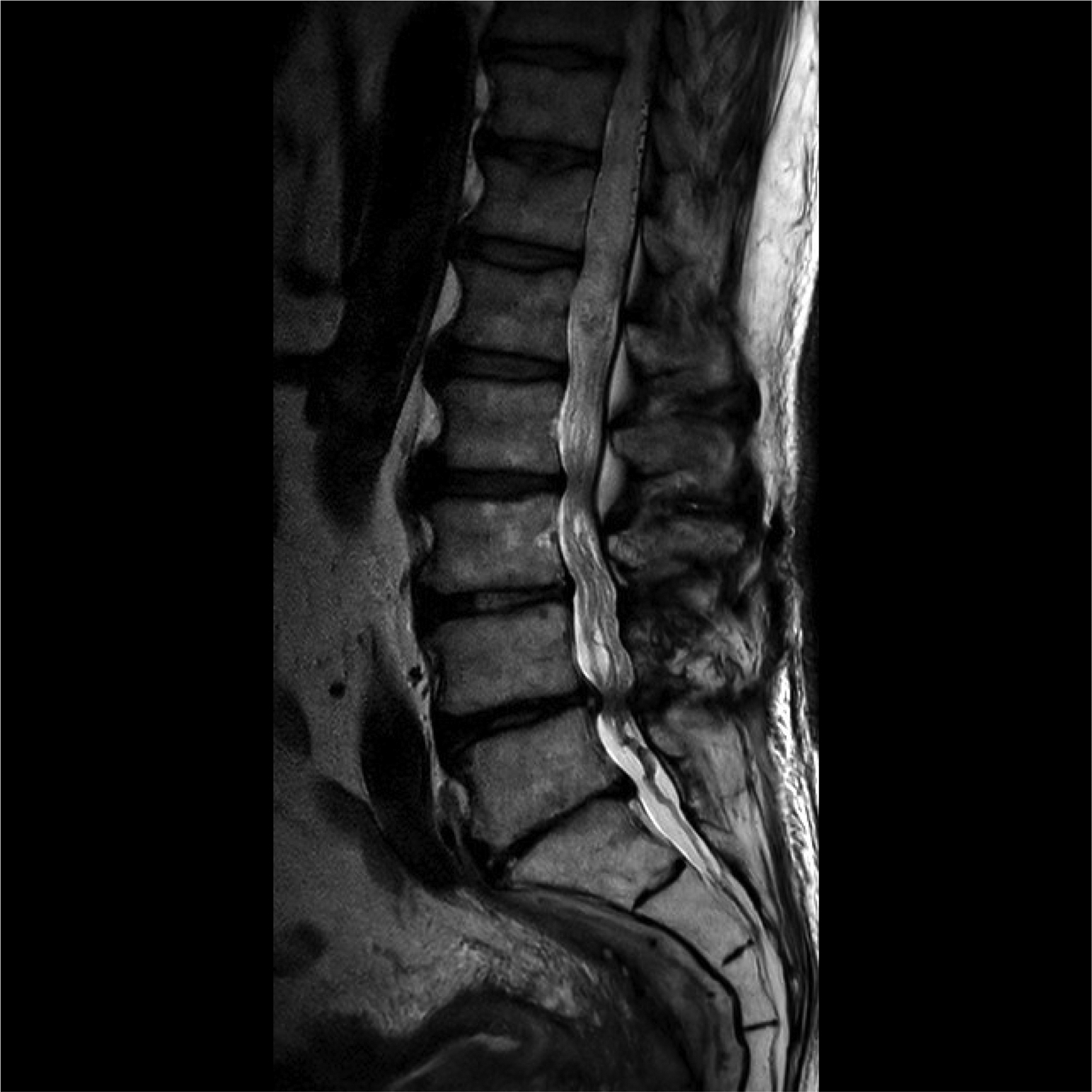
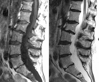
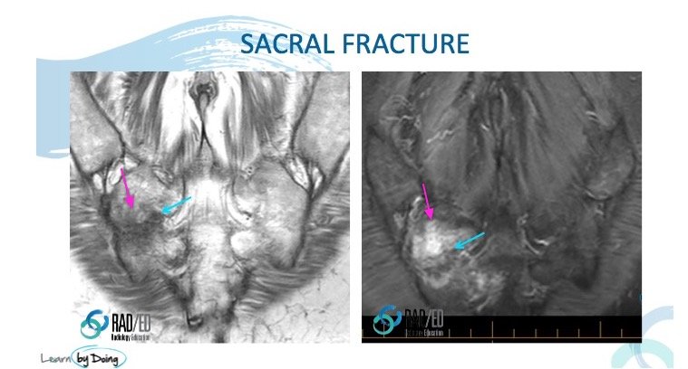
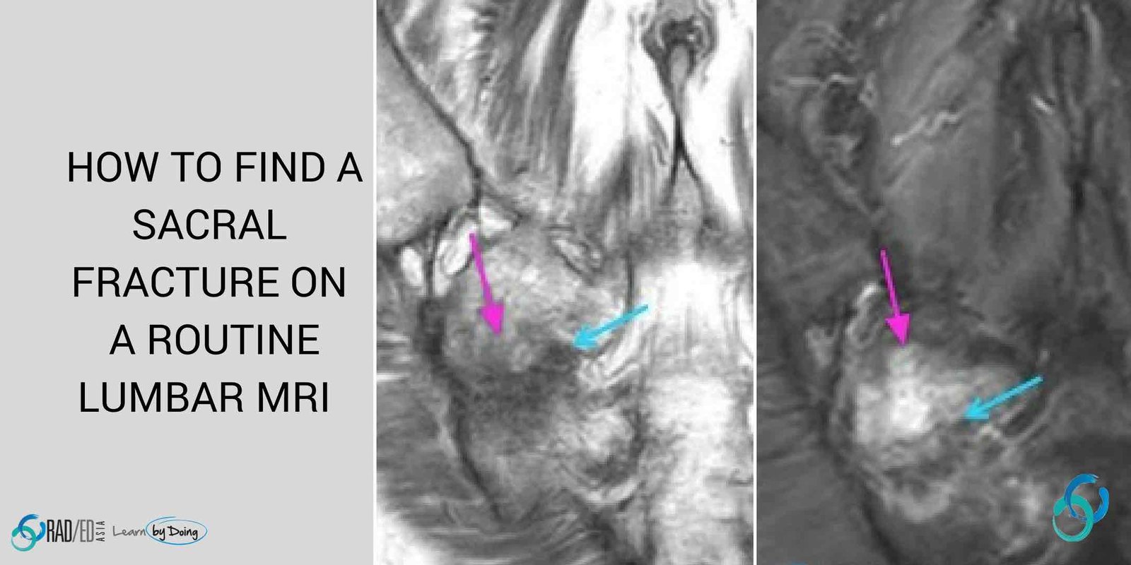
There are 2 things you can do to find a sacral fracture which may be the cause of the. The lumbar spine includes five consecutive vertebrae which are separated by intervertebral discs. Answer 1 of 5. B one marrow signal abnormality in the spine and sacrum is a common sometimes unexpected finding on MRI and it can be a source of diagnostic dilemma to radi-ologists who interpret these examinations. Lumbar MRI will show from the lower thoracic spine t10 or so down to the sacrum and sacroiliac joints. The SIJ should be imaged only if significant clinical findings are demonstrated.
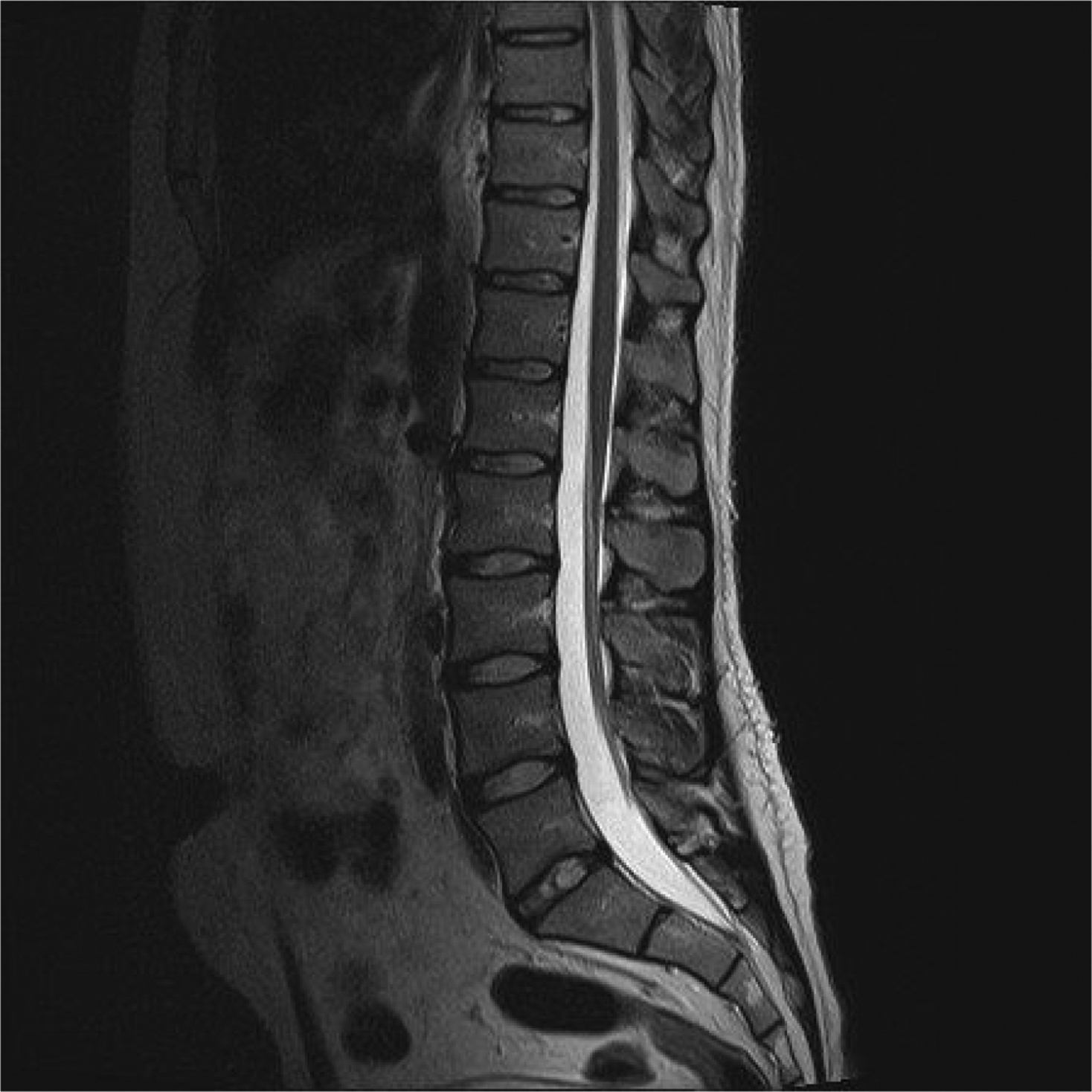
Lumbar Spine 72148 - wo contrast 72149 - wcontrast 72158 - wo wcontrast Other MR Studies MRI Sacrum Coccyx charge as MRI Pelvis. To answer your question no. The SI joints might be seen on some Lsp images but probably not sufficiently to be properly evaluated. A lumbar spine MRI can offer your healthcare provider valuable clues about what is causing your back pain and effective ways to help you find relief. People ask while imaging this can you image that. On a standard lumbar spine MRI with axial and sagittal scans the sacrum is not specifically imaged but sacral fractures can be a source of back pain.
Answer 1 of 4. Diagnostic imaging studies such as x-rays CT scans and MRI scans may show degenerative changes in the SI joints. Cover L4-5 to tip of the coccyx through SI Joints and Sacrum.
Lumbosacral Spine Mri Radiology Key
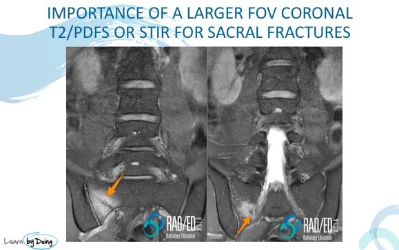
How To Find A Sacral Fracture On A Lumbar Mri Radedasia
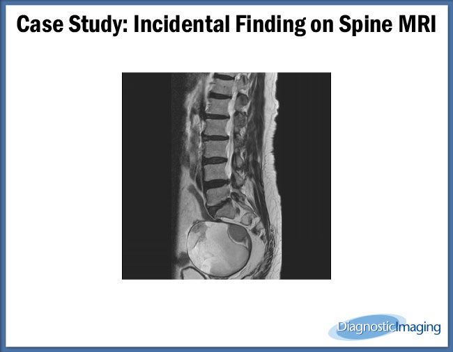
Incidental Finding On Spine Mri
Would An Mri Scan Of An Ls Spine Include An Si Joint Quora
Mr Lumbosacral Plexus Wo Or Wwo Msk Protocol Ohsu
Sacral Insufficiency Fractures Radsource

Lumbar Lumbosacral Spine Mri Planning And Protocols Indications For Mri Lumbar Spine Scan
How To Find A Sacral Fracture On A Lumbar Mri Radedasia

Patient 5 Mri Of Spine Showing Hypoplastic Sacrum Spinal Cord Download Scientific Diagram

How To Find A Sacral Fracture On A Lumbar Mri Radedasia

How To Read And Recognize Normal And Abnormal Lumbar Mri Results

Lumbar Sacral Spinal Mri T2w A Sagittal And B Corona Views Download Scientific Diagram
How To Find A Sacral Fracture On A Lumbar Mri Radedasia
Sacral Cyst Radiology Case Radiopaedia Org
Posting Komentar untuk "Does Lumbar Mri Include Sacrum"