Histology Of The Heart Pdf
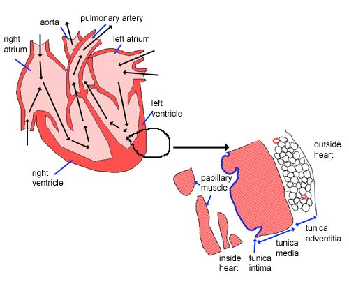
The layers of the heart conform to the basic pattern seen in the histology of other tubular structure except that the muscle layer dominates. The middle layer is the myocardium and the innermost layer is the endocardium which originated from mesothelial cells of the outflow tract.
:watermark(/images/watermark_5000_10percent.png,0,0,0):watermark(/images/logo_url.png,-10,-10,0):format(jpeg)/images/overview_image/1809/Pluv5iH5WLGdFonEHMg_heart-2_english.jpg)
Heart Histology Cells And Layers Kenhub
Structure The heart is divided into 2 sides by the septum.

Histology of the heart pdf. Is in contact with cardiac muscle and contains small vessels nerves and Purkinje Fibers. There are 4 chambers in the heart. PDF On Jan 1 2006 Mohamed Fath El-Bab published Histology II Find read and cite all the research you need on ResearchGate. The heart is an organ composed of muscle nervous connective and epithelial tissues. The erythrocytes are promptly taken up by resident alveolar macrophages. _ NOVEMBER 28 2021 Date.
INTRODUCTION Histology is a word deriving from old greek histos- cloth tissue. These three layers of the heart are embryologically equivalent to the three layers of blood vessels. Logos- word speech meaning science of tissues. The underlying connective tissue layers Subendocardium. ACTIVITY SHEET ACTIVITY 18. Face of the heart embedded in various amounts of subepi- cardial fat.
Histology of Cardiovascular System Histologi - Free download as Powerpoint Presentation ppt PDF File pdf Text File txt or view presentation slides online. Histologi sistem kardiovaskular kuliah umum fk usu 2010. Intima media and adventitia. PDF The heart structure of the spotted scat Scatophagus argus was observed from the Paknam Pranburi Estuary Thailand using histological and. Histology of the Cardiovascular System. Related Books Free with a 30 day trial from Scribd.
In an average persons life the heart will contract about 25 billion times. The increase in pressure of the blood in the pulmonary vasculature results in erythrocytes passing into the alveolar septum. Slideshow is from the University of Michigan Medical Schools M1 Cardiovascular Respiratory sequence. The intima is the inner layer lining the vessel lumen. Download to read offline. Histology of blood vessels The walls of arteries and veins are composed of endothelial cells smooth muscle cells and extracellular matrix including collagen and elastin.
The wall of the heart separates into the following layers. It creates the force that starts the movement of the blood within blood vessels. It provides the researcher with microscopic details of tissues and organs of the human body. Its surrounded by a liquid filled membrane called the pericardium. The endocardium is synonymous with endothelium a single layer of squamous epithelium lying on the basement membrane. This HE stained section provides another good histological study of the heart including part of the aortic valve.
Between the endocardium and myocardium is a layer of variable thickness called the subendocardial layer SEn containing small nerves and in the ventricles the conducting Purkinje fibers P of the subendocardial conducting network. The outermost layer is the epicardium which is derived from the proepicardium from the septum transversum. The heart is a muscular organ that functions as a pump. The cardiovascular system there-. Note the histology of the semilunar valve and the wall of the aorta View Image at the root of the aorta both of which may be seen in close association with the fibrous cardiac skeleton. File Type PDF Histology Of Nervous Tissue Exercise 17 group of different kinds of tissues working together to perform a particular activity.
Tunica adventitia tunica media and tunica intima respectively. HISTOLOGY OF THE HEART JOYLYN L. In heart failure the hearts inability to move blood efficiently results in congestion of the lungs. Then to the larger veins. PDF Junqueiras Basic Histology Text and Atlas 14th Nervous tissue is composed of two types of cells neurons and glial. Approximately 7000 L of blood is pumped by the heart every day.
Epithelium forms internal or external linings of organs and glands specialized for lubrication resisting abrasion water-proofing absorption andor. 44 Anatomy and histology of the heart 67 45 Anatomy and histology of the blood vessels 69 References 72 Chapter 5 Urinary system 75 Elizabeth McInnes 51 Background and development 75 52 Sampling techniques 76 53 Artefacts 76 54 Background lesions 77 55 Anatomy and histology 79 References 85 Chapter 6 Reproductive system 87 Cheryl L. The endocardium En is a thin layer of connective tissue lined by simple squamous endothelium. Blood flow throughout the body begins its return to the heart when the capillaries return blood to the venules and. . Histology of Normal Tissues.
Check the heart slide 102. 27 The Heart and Blood Vesselsnotebook Robert Cummins 9 January 25 2013 The Heart The Heart is made of cardiac muscle that never tires. Epicardium myocardium and endocardium. The thick middle layer of the heart the myocardium m - ō -kar d ē - ŭ m is composed of cardiac muscle cells and is responsible for contrac-tion of the heart chambers. Portions of the epicardial coronary arteries may dip into the myocardium mural artery or tunneled ar- tery and be covered for a variable length 1 to several mm5 by ventricular muscle myocardial bridge Fig. The heart wall has three layers.
Histology of heart disease Qatar Cardiovascular Research Centre. The endothelial lining made of Simple squamous epithelium 2. The endocardium is the inner layer of the heart wall and consists. Endothelium is continous throughout. Histology of liver by aravindh dpi Aravindh Dpi. Find read and cite all.
View HISTOLOGY OF THE HEART AMORONIO JOYLYN 1Bpdf from MLS 1B at University of San Agustin. View additional course materials on OpenMichigan. The heart contains three basic layers similar to those seen in arteries and veins. These are arranged into three concentric layers. The smooth inner surface of the heart chambers is the endocardium en-d ō -kar d ē - ŭ m which consists of simple squamous epithelium over a layer of connective tissue. Heart Failure Cells.
The adventitia is the outer layer of the blood vessel. Tubular structures in the body have a basic structural makeup of an inner layer lined with an epithelial layer abutting the lumen a middle functional layer and an outer protective layer or skin. A double-layer fluid-filled sac known as the pericardium surrounds the heart. Histology of the Heart.
:watermark(/images/watermark_only.png,0,0,0):watermark(/images/logo_url.png,-10,-10,0):format(jpeg)/images/anatomy_term/cardiomyocytes/spYZYp8deRGix6b4SaOnaA_cardiac_muscle_cell.png)
Heart Histology Cells And Layers Kenhub

Mammal Spinal Cord Transverse Section 64x Spinal Cord Mammals Mammals Nervous System Other Systems C Spinal Cord Histology Slides Tissue Biology

Wheater S Functional Histology Free Reading Online E Book Study Smarter

Histology Text Atlas Pdf Am Medicine Medical Textbooks Text Medical Studies

Junqueira S Basic Histology Text And Atlas 15th Edition Pdf Ebook Pdf Digital Textbooks Online Textbook
:watermark(/images/watermark_only.png,0,0,0):watermark(/images/logo_url.png,-10,-10,0):format(jpeg)/images/anatomy_term/cardiac-muscle/5nMjz5Lh5qLyJauAM4IV6A_cardiac_muscle_01.png)
Heart Histology Cells And Layers Kenhub

Lever S Histopathology Of The Skin 11th Edition Pdf Free Pdf Epub Medical Books Ebook Pdf Ebook Medical Textbooks
:watermark(/images/watermark_only.png,0,0,0):watermark(/images/logo_url.png,-10,-10,0):format(jpeg)/images/anatomy_term/heart/dc5loKa5NCrT6VxFUV6cw_Heart.png)
Heart Histology Cells And Layers Kenhub
:watermark(/images/watermark_only.png,0,0,0):watermark(/images/logo_url.png,-10,-10,0):format(jpeg)/images/anatomy_term/cardiomyocyte-nuclei/cRDUrKyJf4oHcXoKnGU6UQ_Nuclei_of_Cardiomyocytes.png)
Heart Histology Cells And Layers Kenhub

Cardiomyopathy Pathology Hypertrophic Cardiomyopathy Heart Conditions Pathology

Mebooksfree Com Free Medical Dr Book Medical Textbooks

Desmosomes And Gap Junctions In Cardiac Muscle Adjacent Muscle Fibers Gap Junctions Intercalated Discs Desmoso Gap Junction Cardiac Muscle Cell Biology Notes

Circulatory System The Histology Guide
Posting Komentar untuk "Histology Of The Heart Pdf"