Focal Calcification In Brain
Research indicates that increased output of the basal ganglia inhibits thalamocortical projection neurons. Atherosclerosis is a pattern of the disease arteriosclerosis in which the wall of the artery develops abnormalities called lesionsThese lesions may lead to narrowing due to the buildup of atheromatous plaque.
Calcified Cerebral Emboli American Journal Of Neuroradiology

The risk factors for cognitive decline were seizure history2 years adjusted odds ratio aOR 3829 95 confidence interval CI.

Focal calcification in brain. Microcalcification is a common feature of both invasive and in situ malignancy. Portable chest X-ray in coronavirus disease-19 COVID-19. At onset there are usually no symptoms but if they develop symptoms generally begin around middle age. A complex partial seizure affects a larger part of the hemisphere and the person. Which assessment findings are the nurse likely to note. Its presence was identified in a wide range of species including human ox sheep horse donkey monkey cow gerbil rat guinea pig chicken and turkey 156.
1970-48194 p 0013 age at onset 1 year aOR 2903 95 CI. The journals editor Yasmin Khakoo MD FAAN in conjunction with the. In simple partial seizures the person remains conscious. 2- Calcification of long bones 3- Pathologic fractures. Patient with a large focus of calcific tendinitis in the supraspinatus tendon. The series is edited and supervised by our associated editor Dr.
1230-6514 p 0013 brain calcification aOR 2375. Similar to CT a systematic approach is best when interpreting MRI brain. MRIs of the brain can be intimidating at first sight because of all the different sequences and parameters. They usually start in the temporal lobe. G9389 is a billablespecific ICD-10-CM code that can be used to. Andrew J Evans Jonathan J James in Breast Pathology 2006.
Brain and Development ISSN 0387-7604 is the Official Journal of the Japanese Society of Child Neurology and is aimed to promote clinical child neurology and developmental neuroscience. You have selected a link to a website operated by a third party. A Axial contrast-enhanced CT scan shows an enhancing vascular lesion in the left parasagittal frontal lobe with internal focal isoattenuating areas representing normal brain parenchyma interspersed within the nidus. People with Sturge-Weber syndrome have varying levels of cognitive function from. When severe it can result in coronary artery disease. The typical appearance is that of small focal globs of amorphous calcification usually seen around the supraspinatus tendon.
1810-9021 p 0008 bilateral leptomeningeal angiomas aOR 3173 95 CI. When these tumors grow inside the brain it increases intra cranial pressure which can cause bran damage and may be even life threatening. Brain death in a vaccinated patient with COVID-19 infection. The journal is devoted to publishing Review Articles Full Length Original Papers Case Reports and Letters to the Editor in the field of Child Neurology and related sciences. However the same general principles of CT head interpretation apply as. Pineal calcification synonyms include corpora arenacea acervuli brain sand psammoma bodies and pineal concretions was observed as early as in 1653 in humans.
Cognitive Neuroscience and Neuroimaging and Biological Psychiatry. Basal ganglia disease is a group of physical problems that occur when the group of nuclei in the brain known as the basal ganglia fail to properly suppress unwanted movements or to properly prime upper motor neuron circuits to initiate motor function. Find a Network Provider in your area. Other specified disorders of brain. 350 mA kvp 120 kV convolution kernel H20s soft tissue window convolution kernel H60s bone window resolution 045 045 5 mm 3 and field-of-view 230 mm. A simple partial seizure can be a precursor to a larger seizure and then it is called an aura.
The features that suggest calcifications are malignant are clustering pleomorphism calcifications of different sizes density and shapes the presence of rod- and branching. Could monochromatic X-rays revolutionize breast imaging. GeneReviews an international point-of-care resource for busy clinicians provides clinically relevant and medically actionable information for inherited conditions in a standardized journal-style format covering diagnosis management and genetic counseling for patients and their familiesEach chapter in GeneReviews is written by one or more experts on the specific. Skull which encloses the brain is very rigid any growth inside this restricted place can cause problems. CT Examination The CT acquisition parameters were as follows. Mutations in PDGFRB which encodes PDGFRβ in pericytes lead to BBB breakdown and cause a primary familial brain calcification characterized by early-onset microvascular calcification in basal.
A client diagnosed with a brain tumor is exhibiting focal symptoms. Calcific tendenitis is commonly seen about the shoulder. Companion titles include Biological Psychiatry. Histopathology confirmed a WHO grade 3 anaplastic oligodendroglioma with focal calcification. We are proud to present the latest paper of our ongoing series New Trends in Breast Imaging. Proliferative type brain AVM in a 27-year-old woman who presented with a 6-year history of headaches and seizures.
Eko-jenik in ultrasonography giving rise to reflections echoes of ultrasound waves. Biological Psychiatry founded in 1969 is an official journal of the Society of Biological Psychiatry and the first in the Biological Psychiatry family of journals. Matthias Dietzel MD MHBA Erlangen Germany In their groundbreaking work Michael Fishman PhD Boston University USA and Madan Rehani. Physiologic Calcification Brain Commonly affects GP more than putamen Seen as normal variant in aging brain Typically in patients 30 years Neurofibromatosis Type 1 Focal areas of increased signal intensity FASI characteristic T2 hyperintense FASI occur in deep gray nuclei GP most common. Our doctors define difficult medical language in easy-to-understand explanations of. The seizures usually involve only one side of the brain focal seizures during which the port-wine birthmark may darken and individuals may lose consciousness.
Partial seizures - also called focal seizures - are seizures which affect only a part of the brain at onset. Therefore you are about to leave the Blue Cross Blue Shield of Mississippi website and enter another website not operated by Blue Cross Blue Shield of Mississippi. Brain Tumor A Brain Tumor is a collection or mass of abnormal cells in Brain. 2016 2017 2018 2019 2020 2021 2022 BillableSpecific Code. These calcifications are in the form of a thick paste of hydroxyapatite crystals. Pediatric Neurology publishes timely peer-reviewed clinical and research articles covering all aspects of the developing nervous systemPediatric Neurology features up-to-the-minute publication of the latest advances in the diagnosis management and treatment of pediatric neurologic disorders.
What type of brain tumor has the potential to cause this mans health problem. Global Open ScienceThe Societys purpose is to promote excellence in scientific research and education in. 1- Acoustic neuroma 2- Meningioma 3- Pituitary adenoma 4- Angioma.
Calcified Cerebral Emboli American Journal Of Neuroradiology


Brain Stones Large Solid Intracranial Calcifications Are More Common Than Previously Thought And While Mri Sequence Nuclear Medicine Mri Medical Imaging
Calcified Neurocysticercosis Lesions Trigger Symptomatic Inflammation During Antiparasitic Therapy American Journal Of Neuroradiology


Porencephaly Radiology Case Radiopaedia Org Radiology Radiology Imaging Brain Images

Neurocysticercosis Radiology Radiology Imaging Brain Images

Multi Focal Calcification Was Identified In The Cerebral Cortex Download Scientific Diagram

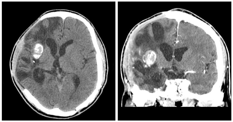
Adamantinomatous Craniopharyngioma Ct Brain Demonstrates A Large Suprasellar Predominantly Cystic Mass With Peripheral Calcification

Multi Focal Calcification Was Identified In The Cerebral Cortex Download Scientific Diagram

An Epigenetic Cause Of Seizures And Brain Calcification Pseudohypoparathyroidism The Lancet
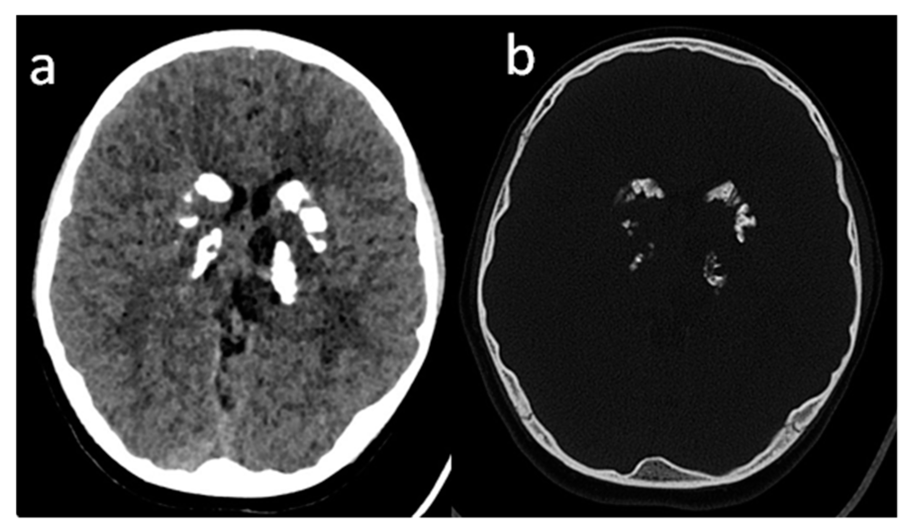
Primary Familial Brain Calcification A 30 Year Old Male With Primary Download Scientific Diagram
Calcified Neurocysticercosis Lesions Trigger Symptomatic Inflammation During Antiparasitic Therapy American Journal Of Neuroradiology

Brain Sciences Free Full Text Leukoencephalopathy With Calcifications And Cysts The First Polish Patient With Labrune Syndrome Html
Intracranial Calcification Caused By A Brain Abscess A Rare Cause Of Intracranial Calcification

A Brain Ct Revealed Marked Intracerebral Calcification In The Bilateral Download Scientific Diagram
Posting Komentar untuk "Focal Calcification In Brain"