Lumbar Mri Feet First
The Boston Scientific Spinal Cord Stimulator Systems are indicated as an aid in the management of chronic intractable pain of the trunk andor limbs including unilateral or bilateral pain associated with the following. Once you have completed lumbar flexion in supine it is time to perform the bending exercise for your stenosis in a seated position.

Lumbar Lumbosacral Spine Mri Planning And Protocols Indications For Mri Lumbar Spine Scan
Lumbar spinal stenosis LSS is a medical condition in which the spinal canal narrows and compresses the nerves and blood vessels at the level of the lumbar vertebraeSpinal stenosis may also affect the cervical or thoracic region in which case it is known as cervical spinal stenosis or thoracic spinal stenosis.

Lumbar mri feet first. The rule in health care is the same as in money. Slowly bend yourself forward and reach towards the floor. Clinical Biomechanics is an international multidisciplinary journal of biomechanics with a focus on medical and clinical applications of new knowledge in the field. MRI images were independently evaluated by two neurosurgeons with consensus one with more than 6 years of experience and a special interest in spine surgery and one with more than 22 years of. Once inserted the spacers arms open around the spinous processes the bumps you can feel in your spine to help make proper space for the affected nerves. The first grade is known as a lumbar disc protrusion.
Sufficient documentation such as history and physical notes laboratory results signs and symptoms of the disease to warrant the diagnostic test and to support the claim of reasonable. Mild retrolisthesis of L4 and L5 noted. Complications and risks of lumbar laminectomy include nerve damage bleeding infection and blood clots. Anon993988 January 4 2016. Aetna considers magnetic resonance imaging MRI studies of the knee medically necessary when any of the following criteria is met. Similarly kyphosis historically refers to abnormal convex curvature of the spine.
Lumbar spondylosis is a degenerative condition that develops gradually over time being more common in older individuals. A lumbar laminectomy is a surgery that removes most of the bony arch of a vertebra to treat lower back pain. Lumbar spinal stenosis can cause pain in the low back or buttocks. Care properly for your grade one lumbar disc protrusion and you will not end up with a grade two. Or Persistent knee painswelling andor instability giving way not associated with an injury and not responding to at least 3 weeks of. With world-class physicians on staff the newest and most advanced technology and a patient experience pathway that is unrivaled in its efficiency and pedagogy of care Deuk Spine Institute has performed thousands of procedures and achieves a 95 success rate in elimination of pain.
Why do they call it lumbar spondylosis if as doctors have told me it is osteoarthritis. The science of biomechanics helps explain the causes of cell tissue organ and body system disorders and supports clinicians in the diagnosis prognosis and evaluation of treatment methods and. The soles of the foot receive their nerve supply from the first sacral root S1. This section of the website will explain how to plan for an MRI wrist scans protocols for MRI wrist how to position for MRI wrist and indications for MRI wrist. To determine the cause of this uncomfortable feeling in your feet see your primary care physician who can take your medical history examine you and perform the necessary tests. Osteoporotic spinal fractures are unique in that they may occur without apparent trauma.
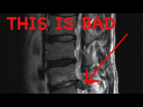
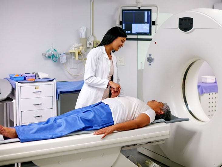
MRI magnetic resonance imaging This scan can reveal problems with your soft tissues. Accurate coding information must be provided with claims to differentiate CT andor MRI scans from other radiology services and to make coverage determinations. 3The conus medullaris is at T12-L1 level 4The lumbar vertebral bodies show normal stature and marrow signal characteristics. Any injury that changes. And ultimately progressing to grade four. MRI lumbo -sacral spine Report 1Numbering of vertebrae done by counting from C2 downwards 2Exaggerated lumbar lordosis is noted.
For descriptive purposes lumbosacral plexus is normally divided into three main parts lumbar plexuses sacral plexuses and pudendal plexuses. Phase direction in the coronal scans must be head to feet with 100 over. However the terms lordosis and lordotic are also used to refer to the normal inward curvature of the lumbar and cervical regions of the human spine. MRI scan myelogram CT scan or EMG. Degenerative Disc Disease Treatment. Herniated lumbar disc.
Suggested sequence parameters and planning. To perform lumbar flexion in sitting. It consists of five vertebrae known as L1 - L5. I am a 29 year old woman and like many of you I have suffered with the effects of lumbar spondylosis for years. A small spacer is placed inside the spine without removal of any nearby bone or tissue. This condition can also be referred to as spinal osteoarthritisIt occurs.
The lumbar spine is located in the lower back below the cervical and thoracic sections of the spine. If symptoms continue surgery may be recommended. An example of lumbar low back degenerative disc disease in an imaging study. Modic type 2 changes noted at multiple levels. These lumbar vertebrae or lumbar bones contain spinal cord tissue and nerves which control communication between the. The normal outward convex curvature in the thoracic and sacral.
Register the patient in the scanner as head first supine. Failed back surgery syndrome Complex Regional Pain Syndrome CRPS Types I and II intractable low back pain and leg pain. Sit in a firm chair with both feet on the floor. Based on the results you may be referred to a. Lumbar laminectomy is often performed in combination with other types of back surgery such as lumbar laminotomy and discectomy. This is a common root to be compressed in spinal stenosis.
If the narrowing is substantial it can cause compression of the spinal cord or spinal nerves. Look after the pennies and the pounds will look after themselves. Osteoporosis is the underlying cause of many lumbar fractures especially in postmenopausal women. Hold the fully bent position for 2 seconds. Lordosis is historically defined as an abnormal inward curvature of the lumbar spine. Most people improve in 6 weeks and return to normal activity.
Fractures of lumbar vertebrae occur in the setting of either severe trauma or pathologic weakening of the bone see image R L4 compression fracture. Once the subject was entered in the study multiplanar MRI was done from the first lumbar to the first sacral vertebra with a 15-tesla imaging system. Treatment with rest pain medication spinal injections and physical therapy is the first step to recovery. Detection staging and post-treatment evaluation of tumor of the knee. Lumbar plexus is divided into six main braches Iliohypogastric Ilioinguinal genitofemoral lateral femoral cutaneous Femoral Obturator and accessory obturator. I was 16 when it first starting causing problems.
It can also show if your discs have shrunk if your spinal canal has narrowed or if your spinal discs are damaged. Lumbar spinal stenosis is a condition where the spinal canal central stenosis or one or more of the lumbar vertebral foramina foraminallateral stenosis becomes narrowed.
Open Mri Of Delafield Llc Lumbar Spine Mri Comfortable Feet First Position And You Can Look Out The Window During The Exam Facebook


Pin On Spinal Injuries

How To Read And Recognize Normal And Abnormal Lumbar Mri Results
How To Read And Recognize Normal And Abnormal Lumbar Mri Results

Back Pain And Normal Mri Is It Possible Advanced Pain Management Center Interventional Pain Management Physician

Lumbar Lumbosacral Spine Mri Planning And Protocols Indications For Mri Lumbar Spine Scan

Do I Really Need An X Ray Or Mri For Lower Back Pain

What The Doctor Is Looking For In A Spine Mri Advanced Bone Joint
How To Read A Spine Mri Youtube
Lumbar Mri Scan Purpose Procedure And Risks

Diagnostic Imaging Spine Mri Cervical Thoracic Lumbar

Color Mri Of The Lumbar Spine Low Back Showing A Normal Lumbar Spine And One With Disc Herniations Created By Medical Media Imag Disk Herniation Mri Anatomy

How To Read An Mri Scan Of The Lumbar Spine Lower Back Part 1 First Look Mri Youtube

Lumbar Spine Magnetic Resonance Imaging Mri Images Demonstrating An Download Scientific Diagram
Posting Komentar untuk "Lumbar Mri Feet First"