Capillary Telangiectasia Vs Cavernoma
Capillary telangiectasias represent localized collections of dilated capillary-like vessels interspersed within normal brain. Spider telangiectasis is an acquired vascular malformation.

Cerebral Cavernous Malformation What A Practicing Clinician Should Know Mayo Clinic Proceedings
It occurs because of the failure of a tiny muscle restricting the size of an arteriole.

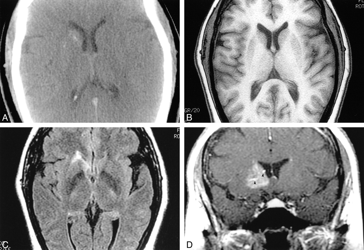
Capillary telangiectasia vs cavernoma. Capillary telangiectasias CTSs are small areas of abnormally dilated. McCormick PW et al Cerebellar hemorrhage associated with capillary telangiectasia and venous angioma. Bleeding episodes are less deadly than with AVMs and surgery is less risky. Axial FLAIR reveals a lobulated lesion with a low signal rim suggestive of hemosiderin in keeping with a cavernomaA subtle flow void is seen extending to the cortical surface suggestive of an associated venous anomaly. An angioma or haemangioma is a benign tumour formed by the dilation of blood vessels or the formation of new ones by the proliferation of endothelial cells. Venous angioma in pontine area.
Infantile haemangioma superficial deep or mixed. Critics claim the distinction is arbitrary but have been unable t. Cavernous angioma ie cavernous hemangioma cavernoma has an increased risk of bleeding if it grows in size. It is involved in relaying signals for respiration sleep. Up to 10 cash back Cavernomas may be calcified and have a typically pop-corn like appearance on MRI. The condition has generally been found in 3 regions of the central nervous system.
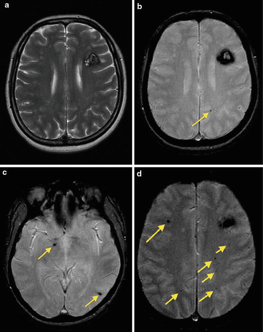
Thin walled dilated vascular channels. Learn to diagnose and treat Telangiectasia immediately. Only scarce case reports have described the utility of SWI in. DVA venous angioma is a collection of enlarged veins. They are reported as scattered dots or stippled in the vessel wall or adjacent brain parenchyma commonly in non-hemorrhagic lesions. These findings are considered.
Capillary telangiectasias are being recognized with increasing frequency on MR imaging studies. BCT is a benign entity whose appearance on conventional MR imaging makes its differentiation from neoplastic inflammatory or subacute ischemic disease challenging. May be caused by a more serious disease called hereditary hemorrhagic telangiectasi. I have been told a have probably a small central pontine capillary telangiectasia vs. Due to recurrent bleeding tuberance carvernomas are surrounded by a pseudocapsule of common gliotic brain that is stained with hemosiderin Fig. Spider telangiectasis may arise spontaneously or may be induced by.
Arteriovenous venous cavernous and capillary telangiectases. Increased pulsating flow through the vessel the central papule results in the dilatation of distal vessels the red lines. Most are located in the brain stem and show slightly increased signal intensity on T2-weighted images low signal intensity on T2-weighted images reflecting the presence of deoxyhemoglobin and contrast enhancement. Surgical resection is recommended for cavernomas presenting with symptomatic hemorrhage in accessible and non-eloquent locations. Cerebral vascular malformations have traditionally been divided into four categories. These are sometimes syndromic as in PHACE.
May look like petechial hemorrhages. Axial T1 reveals areas of increased signal within the lesion in keeping with methemoglobin suggestive of recent hemorrhage. Most common vascular malformation found at autopsy. Offering a Full Range of the Latest Treatments for Cavernoma. Capillary telangiectasia consist of irregular clusters of dilated capillaries intermixed with normal brain parenchyma and are most often located in the pons1 Based on their relatively common incidental discovery at necropsy in people without overt neurological manifestations brain stem capillary. Calcifications in venous angioma and capillary telangiectasias have been described scarcely.
Lesions larger than capillary teleangiectasia. Ad Telangiectasia is a condition that causes thread-like lines to appear on the skin. Cavernomas and AVMs in particular can be more serious diagnoses as they have the potential to cause debilitating symptoms including the risk of rupture and bleeding into the brain. Learn the best ways to treat Telangiectasia right now. Capillary telangiectasias are composed of multiple thin-walled vascular channels between normal brain parenchyma. A controversy exists about separating the latter two lesions into separate entities.
The Zabramski classification of cerebral cavernomas has been proposed as a way of classifying cerebral cavernous malformations and although not used in clinical practice it is useful in scientific publications that seek to study cavernous malformations. AKA Campbell De Morgan spots. Ad See Doctors with Extensive Experience in Diagnosing Treating Cavernoma. The classification was proposed in 1994 1 and at the time of writing June 2016 remains the most commonly. Venous angioma detected using gadolinium enhancement on MRI. This was detected during my MRI during my MS workup.
In cavernous angioma they are seen in up to 33 of the cases. BACKGROUND AND PURPOSE. Capillary Telangiectasia CTS is a type of vascular malformation of the brain in which clusters of dilated capillaries are formed in some areas of the brain interspersed with normal brain tissue. Lesions which distinguishes cavernomas from capillary telangiectasias10 Macroscopically cavernomas are bluish nodules contain-ing In areas of bleeding at different stages. Proliferates in the first few weeks of life followed by involution later in childhood. SWI is sensitive to susceptibility effects from deoxyhemoglobin with excellent spatial resolution.
A capillary telangiectasia is small lesion made of tiny vessels.
Epos Trade


References In De Novo Formation Of A Cavernous Malformation Of The Brain In The Presence Of A Developmental Venous Anomaly Clinical Radiology
Mr Imaging And Histologic Features Of Capillary Telangiectasia Of The Basal Ganglia American Journal Of Neuroradiology

Pin By Karl D On Neurosurgery Brain Lesions Brain Surgery Neurology

Vascular Malformations Neupsy Key

Giant Symptomatic Mixed Vascular Malformation Containing A Cavernoma Developmental Venous Anomaly And Capillary Telangiectasia In A 19 Month Old Infant Springerlink
Vascular Malformations Avms Venous Angioma Cavernous Malformation Capillary Telangiectasia Flashcards Quizlet

Types Of Vascular Malformations Vascular Veins Syndrome
Neurology And Neuroscience Karger Find Out More About Cerebral Cavernous Malformations Developmental Venous Anomaly And Its Coexistence In This Review Http Ow Ly Osyg50bsy3k Pretty Sara Idiculla European Neurology Facebook


Cerebral Cavernous Malformation What A Practicing Clinician Should Know Mayo Clinic Proceedings

Miscellaneous Vascular Malformations Cavernous Malformations Developmental Venous Anomaly Capillary Telangiectasia Sinus Pericranii Radiology Key

Cerebral Cavernous Malformation What A Practicing Clinician Should Know Mayo Clinic Proceedings
2
Miscellaneous Vascular Malformations Cavernous Malformations Developmental Venous Anomaly Capillary Telangiectasia Sinus Pericranii Radiology Key
Posting Komentar untuk "Capillary Telangiectasia Vs Cavernoma"