Can Nerve Damage Be Detected On An Mri
36 Even though a nerve injury may be evident following clinical and electrodiagnostic testing it is frequently unclear whether the nerve has suffered a neuropraxia axonotmesis or neurotmesis in the early. Women are more vulnerable than men to many of the medical consequences of alcohol use.
Normal Anatomy Of The Sciatic Nerve A Axial T1weighted Mr Image Download Scientific Diagram
At fixed time intervals after administration the contrast agent mixing with the blood will arrive at various sites in the body which can then be scanned.
Can nerve damage be detected on an mri. However a venous puncture may be undetected if aspiration or palpation pressure collapses the venous lumen. Pain that arises from actual or threatened damage to non-neural tissue and is due to the activation of nociceptors nerve endings that detect tissue damage The next definition is also based on this reference. For example alcoholic women develop cirrhosis 5 alcoholinduced damage of the heart muscle ie cardiomyopathy 6 and nerve damage ie peripheral neuropathy 7 after fewer years of heavy drinking than do alcoholic men. One can also monitor nerve recovery during the period of rehabilitation especially from four months when regeneration can be detected. Because acute bacterial meningitis can lead to permanent brain or nerve damage or death within hours treatment is started as soon as possible without waiting for the results of diagnostic tests and often before a spinal tap is done. Pain caused by a lesion or disease of the somatosensory nervous system.
Detect damage to the brain caused by an injury or a stroke. Vascular Puncture Vascular puncture can occur with an axillary block but it usually can be detected. A persons susceptibility to alcoholismrelated brain damage may be associated with his or her age gender drinking history and nutrition as well as with the vulnerability of specific brain regions. The electric current does not come into contact with the patient. We would like to show you a description here but the site wont allow us. 51 52 Moreover MRI is better at imaging the brainstem basal ganglia and thalami.
Nerve damage or trauma tumor inflammation radiation damage compression related to disc disease or entrapment ie. Alcoholism can affect the brain and behavior in a variety of ways and multiple factors can influence these effects. Joel Block Editor-in-Chief of Osteoarthritis and Cartilage would like to recognise the following reviewers and thank them for their contribution to the journal. 1 LBP and 2 radicular symptoms in the lower extremities which in some cases lead to neurogenic. A coma is a deep state of prolonged unconsciousness in which a person cannot be awakened fails to respond normally to painful stimuli light or sound lacks a normal wake-sleep cycle and does not initiate voluntary actions. Low back pain is essentially the modern-day pandemic with chronic low back pain LBP being the second leading cause of adult disability in the United States.
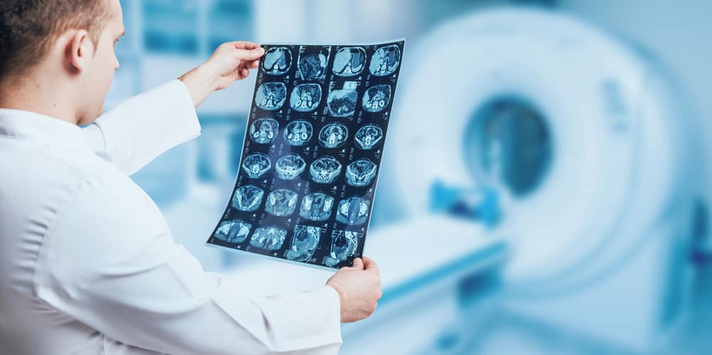
Magnetic resonance imaging MRI is a noninvasive test doctors use to diagnose medical conditions. Tobias Bäuerle Francisco J. To promote equity and diversity among authors reviewers and editors. The symptoms of degenerative lumbar spine disease can broadly be divided into two broad categories. To provide a platform for discussion of current ideas in urologic education patient engagement. Functional magnetic resonance imaging or functional MRI fMRI measures brain activity by detecting changes associated with blood flow.
Pediatric Neurology publishes timely peer-reviewed clinical and research articles covering all aspects of the developing nervous systemPediatric Neurology features up-to-the-minute publication of the latest advances in the diagnosis management and treatment of pediatric neurologic disorders. MRI is superior to CT in detecting axonal injury small areas of contusion and subtle neuronal damage. However magnetic resonance imaging Computed tomography CT and magnetic resonance imaging MRI A doctor can often diagnose a musculoskeletal disorder based on the history and the results of a physical examination. This technique relies on the fact that cerebral blood flow and neuronal activation are coupled. When an area of the brain is in use blood flow to that region also increases. These coils send and receive radio waves producing signals that are detected by the machine.
Comas can be derived by natural causes or can. Scan phases include the arterial portal venous nephrogenic and excretion phases fig. At field strengths of 7 T and higher MRI can detect not only hydrogen nuclei but also the nuclei of heavier elements such as sodium potassium phosphorus and fluorine which have a much lower. MRI demonstrating promise in both diagnosing and monitoring injury especially in the surgical setting. The journals editor Yasmin Khakoo MD FAAN in conjunction. Blanco Jeroen Geurts Tariq M Haqqi Satoshi Kubota.
Thoracic outlet piriformis. MR neurography can identify nerve discontinuity of a nerve but over 50 of high-grade nerve transections have minimal to no gap present. EEG also has some characteristics that compare favorably with behavioral testing. Laboratory tests imaging tests or other. Aviation History magazine is an authoritative in-depth history of world aviation from its origins to the Space Age. Diffusion tensor imaging DTI a type of MR can quantify axon density and myelin thickness.
The mission of Urology the Gold Journal is to provide practical timely and relevant clinical and scientific information to physicians and researchers practicing the art of urology worldwide. Coma patients exhibit a complete absence of wakefulness and are unable to consciously feel speak or move. MRN can be performed at any time after nerve injury as opposed to electrodiagnostic studies which usually require a two- to three-week waiting period after abnormalities can be detected. EEG can detect covert processing ie processing that does not require a response EEG can be used in subjects who are incapable of making a motor response Some ERP components can be detected even when the subject is not attending to the. Arthrography can be used to view torn ligaments and fragmented cartilage in the joint. Aviation History offers air enthusiasts the most detailed coverage of the history of manned flight with action-packed stories and illustrations that put the reader in the cockpit with pilots and military Army Navy and Marines aviators to experience aviations greatest dramas.
The formal definition. Thank you to 2020s top reviewers. Additionally enhancements blood supply perfusion of the abdominal organs can be evaluated. Symptoms of nerve damage sensory loss and. A Google ingyenes szolgáltatása azonnal lefordítja a szavakat kifejezéseket és weboldalakat a magyar és több mint 100 további nyelv kombinációjában. 49 50 Studies have shown that CT missed approximately 1020 of abnormalities seen on MRI.
Affects several nerves and is usually accompanied by soreness of the upper arm.
Will Nerve Damage Show Up On An Mri Aica Orthopedics

Acute Optic Nerve Lesions In First Ever Nmosd Related Optic Neuritis Using Conventional Brain Mri A Latin American Multicenter Study Multiple Sclerosis And Related Disorders
Can You See Nerve Damage In An Mri
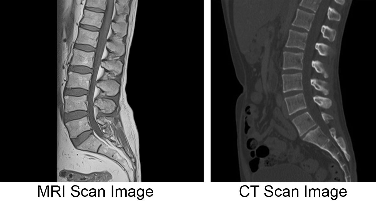
Mri Vs Ct Scan Diagnosing Spine Neck Injuries Degenerative Diseases Joseph Spine Institute

Back Pain And Normal Mri Is It Possible Advanced Pain Management Center Interventional Pain Management Physician
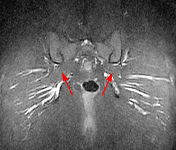
Piriformis Syndrome Radsource
Can You See Nerve Damage In An Mri

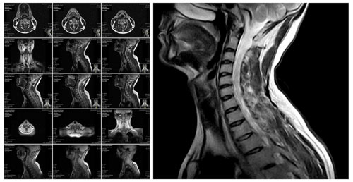
Head And Neck Mri Neurological Specialists P C
Can An Mri Show Optic Nerve Damage Quora
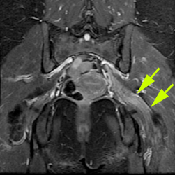
Role Of Mr Neurography For The Diagnosis Of Peripheral Trigeminal Nerve Injuries In Patients With Prior Molar Tooth Extraction American Journal Of Neuroradiology


Magnetic Resonance Imaging For Back Pain Mriplus
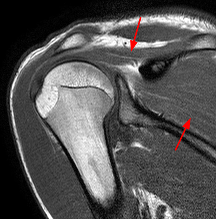
Parsonage Turner Syndrome Radsource
Mr Neurography Mr Imaging Of Peripheral Nerves Pni Ucsf Radiology
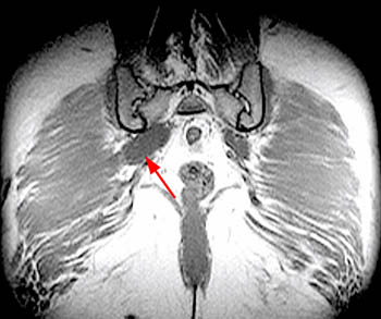
Piriformis Syndrome Radsource
Posting Komentar untuk "Can Nerve Damage Be Detected On An Mri"