Who Classification Of Brain Tumors 2016 Radiology
Characterization of these soft-tissue lesions remains problematic despite advances in imaging. Many cases never produce symptoms.

Colloid Cyst With Hydrocephalus Well Defined Hyperdense Mass At The Foramen Of Monroe Causing Obstructive Hydrocephalus Radiology Brain Images Brain Surgery
Adipocytic tumors fibroblastic or myofibroblastic tumors so-called fibrohistiocytic tumors smooth muscle tumors skeletal.

Who classification of brain tumors 2016 radiology. Claus Lunau Science Photo Library Subarachnoid haemorrhage is the result of a haemorrhage from a cerebral blood. Magnetic resonance imaging MRI contrast agents are categorised according to the following specific features. The new 2016 WHO Classification of Tumors of the Central Nervous System was a paradigm shift. 1 3 Despite this increased recognition it has yet to become an established diagnostic consideration outside of large tertiary referral centers. Benign and malignant breast tumors classification based on region growing and CNN segmentation ESA 2015 Adversarial Deep Structured Nets for Mass Segmentation from Mammograms ISBI 2018 paper Improved Breast Mass Segmentation in Mammograms with Conditional Residual U-net MICCAI 2018 paper. In this classification three new entities seromucinous hamartoma NUT carcinoma and biphenotypic sinonasal sarcoma were included while the total number of tumors has been.
Autoimmune encephalitis is an important cause of new-onset altered mental status the scope of which has only recently begun to be recognized in the medical literature. The World Health Organization WHO 2017 classification of head and neck tumors has been just published and has reorganized tumors of the nasal cavity and paranasal sinuses. By systematically using clinical history lesion location mineralization on radiographs and signal intensity characteristics on magnetic resonance images one can a determine the. Knowing the location of a hemorrhage is often the key to the differential diagnosis especially in non-traumatic bleeding. MR images of the patient revealed a non-contrasting well-circumscribed lesion lodged in the pituitary gland. Algorithmic and Computer-Based Approaches Studies in Computational Intelligence 1st ed.
Non-invasive Treatment of Solid Tumors 1st ed. Some of the tumors were defined also by their genetic composition as well as their cell morphology. Extent of surgery and WHO grading J Neurosurg 20151224 Neuro Oncol 201618863 Most recently DNA methylation profiling is reported to better predict tumor recurrence and prognosis than the WHO histological classification Lancet Oncol 201718682 Recurrent losses of chromosome 1p 6q 14q 18q and gain of 1q are indicators of poor. In the most recent World Health Organization WHO system for classification of soft tissue tumors which was published in 2002 soft tissue tumors are grouped into nine major categories based on their predominant histologic makeup. Occasionally seizures dementia trouble talking vision problems one. Primary intracranial tumors of the brain structures including meninges are rare with an overall five-year survival rate of 334.
The majority of these agents are either paramagnetic. Academic Radiology publishes original reports of clinical and laboratory investigations in diagnostic imaging the diagnostic use of radioactive isotopes computed tomography positron emission tomography magnetic resonance imaging ultrasound digital subtraction angiography image-guided interventions and related techniques. Symptoms depend on the location and occur as a result of the tumor pressing on nearby tissue. Paranasal CT scans revealed a. Soft-tissue lesions are frequently encountered by radiologists in everyday clinical practice. 2016 Edition Image-Guided Stereotactic Radiosurgery.
The journals editor Yasmin Khakoo MD FAAN in conjunction with the. Radiology department of the University Medical Centre of Utrecht and the Alrijne Hospital in Leiderdorp the Netherlands Publicationdate 2016-01-15 This is an updated version of the 2007 article. Primary tumors of the spine are rare with a reported incidence of 25 to 85 per 100000 people per year They account for less than 5 of new bone tumors diagnosed every year in the United States These tumors exhibit characteristic imaging features that can help in early diagnosis and improved prognosis. Extra-axial hemorrhage - Intracranial extracerebral Subarachnoid hemorrhage is acute bleeding under the arachnoidMost commonly seen in rupture of an aneurysm or as a result of trauma. Case-Oriented Fast Focused Effective Education Imaging Non-Traumatic Abdominal Emergencies in Pediatric Patients 2017 Emergency Radiology of the Chest and Cardiovascular System 2016. The grading of gliomas changed importantly and glioblastoma was now mainly classified according to the status of isocitrate dehydrogenase IDH mutation.
The diagnostic component is based on the 2016 update of the WHO Classification of Tumors of the Central Nervous System and the subsequent recommendations of the Consortium to Inform Molecular and Practical Approaches to CNS Tumour Taxonomy -. High-Precision Medical Imaging in Clinical Applications. Chemical composition including the presence or absence of metal atoms route of administration magnetic properties effect on the magnetic resonance image biodistribution and imaging applications. 1 5 The term autoimmune. Pediatric Neurology publishes timely peer-reviewed clinical and research articles covering all aspects of the developing nervous systemPediatric Neurology features up-to-the-minute publication of the latest advances in the diagnosis management and treatment of pediatric neurologic disorders. A brain and skull cross section showing the different anatomical layers Source.
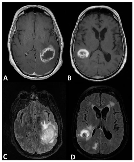
Emergency Radiology Coffee Case Book. A 32-year-old male patient was admitted with complaints of headache and blurred vision. This lesion was intense and sporadically hypointense in T1A imaging and heterogenically hyperintense in T2A sections Fig. Meningioma also known as meningeal tumor is typically a slow-growing tumor that forms from the meninges the membranous layers surrounding the brain and spinal cord.
Update On Brain Tumor Imaging From Anatomy To Physiology American Journal Of Neuroradiology

2
Update On Brain Tumor Imaging From Anatomy To Physiology American Journal Of Neuroradiology

2

Radiology Signs Radiology Radiology Imaging Medical Illustration
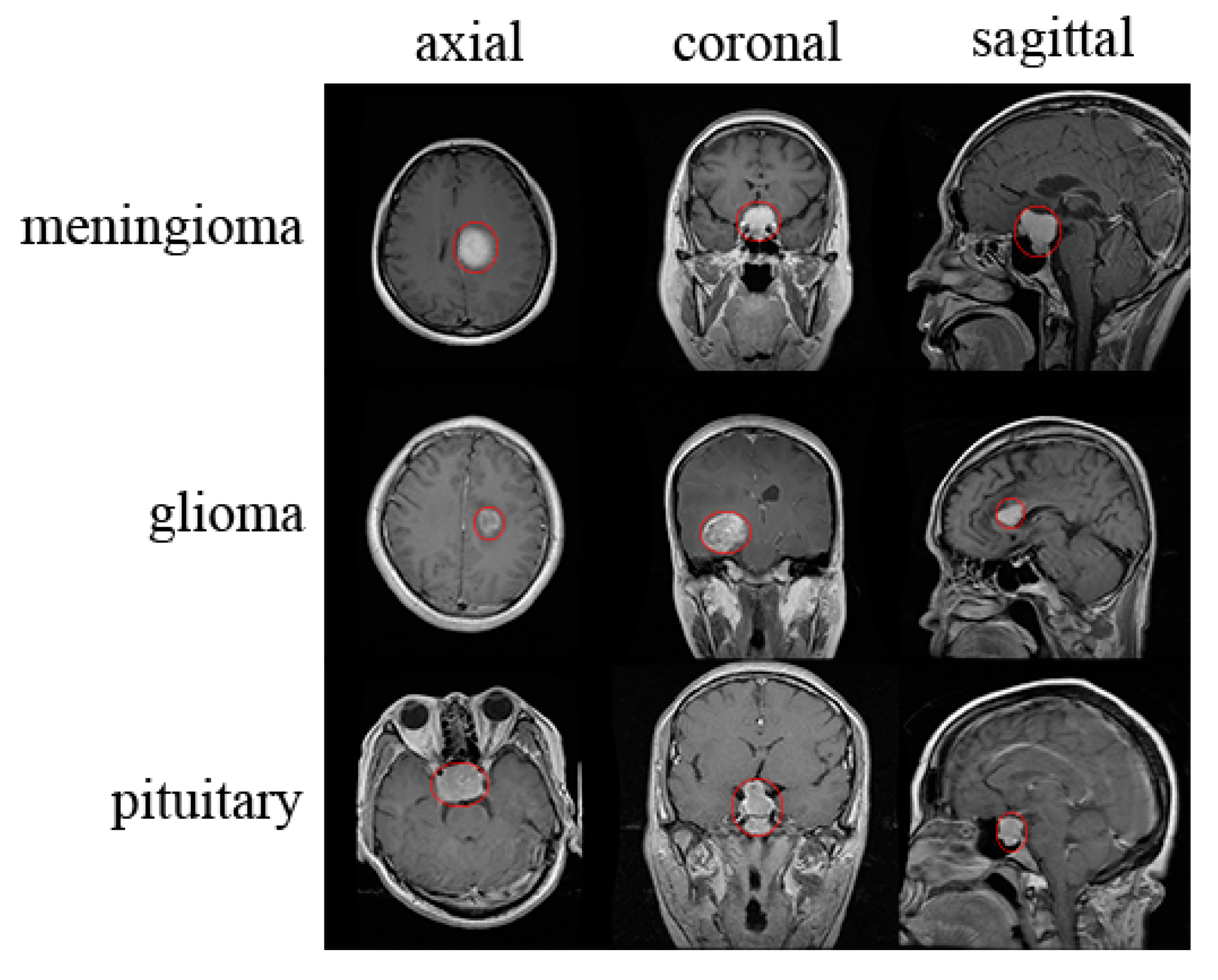
Applied Sciences Free Full Text Classification Of Brain Tumors From Mri Images Using A Convolutional Neural Network Html

The 2016 World Health Organization Classification Of Tumors Of The Central Nervous System A Summary Acta Neuropathologica Central Nervous System Nervous System Tumor
Cancers Free Full Text Differentiating Glioblastomas From Solitary Brain Metastases An Update On The Current Literature Of Advanced Imaging Modalities Html
Pin On Radio Brain
2
2

Pin On Radio Brain
Brain Tumor Types Johns Hopkins Medicine
2
Posting Komentar untuk "Who Classification Of Brain Tumors 2016 Radiology"