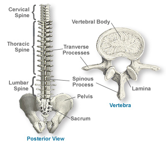
Weight Bearing Portion Of The Vertebra
During a car crash the forearm is slammed against the dashboard. Osteoporosis is a disease of bone in which bone density is reduced which may increase the chance that a person could sustain a vertebral compression fracture with little or no trauma.
First lets take a look at this muscle.

Weight bearing portion of the vertebra. The processes serve as attachment points for various ligaments and muscles that are important to the stability of the spine. The preserved portion of the holotype measures 36 metres 12 ft with only the end of the tail being missing. The deep fibers of the psoas muscle originate on the transverse processes of L1- L5 while the superficial fibers arise from the lateral surfaces of the lumbar vertebra and adjacent intervertebral discs. The length of the tail including the unpreserved portion was later estimated to be 25 metres 82 ft resulting in a total estimated body length of 45 metres 15 ft for the animal though its been argued this is. The proximal surface of the radius articulates with the humeral capitulum which is not as prominent as in the human. Vertebral compression fractures can be caused by osteoporosis trauma and diseases affecting bone pathologic fracture.
The sacrum is a triangular-shaped bone that is thick and wide across its superior base where it is weight bearing and then tapers down to an inferior non-weight bearing apex Figure 10. The total cross-sectional area of the load-bearing calcified portion of the two forearm bones radius and ulna is approximately 24 cm2. Osteoporosis most commonly occurs. The limbs of the horse are structures made of dozens of bones joints muscles tendons and ligaments that support the weight of the equine body. The iliopsoas is compsosed of two separately identifiable parts the psoas and the iliacus. The posterior or back aspect of the body and medial or inside aspects of the pedicle and the anterior or front lamina form a protective.
It is formed by the fusion of five sacral vertebrae a process that. The radius is the medial forearm bone and is the main weight-bearing bone of the antebrachium distally. 6 in the cervical region neck 12 in the thoracic region middle back and 5 in the lumbar region lower back. The intervertebral disc IVD is important in the normal functioning of the spine. They include two apparatuses. Tethyshadros was originally thought to be a dwarf hadrosauroid.
The canine distal radius has distinct facets for articulation with carpal bones providing stability in weight bearing. The suspensory apparatus which carries much of the weight prevents overextension of the joint and absorbs shock and the stay apparatus which locks major joints in the limbs allowing horses. It is a cushion of fibrocartilage and the principal joint between two vertebrae in the spinal column. There are 23 discs in the human spine. The vertebral bodies are the major weight bearing portion of the vertebra. If a portion of wet articular cartilage with all of its layers was separated into the most important individual elements water would provide 6580 of its weight type II collagen fibrils would account for 1020 along with very small percentages of other collagen types and 1015 would be made up primarily of Aggrecan but also.
The arm comes to rest from an initial speed of 80 kmh in 50 ms.
The Vertebral Column Joints Vertebrae Vertebral Structure
Question Video Recognizing The Structures Of A Vertebra Nagwa
Axial Skeleton Vertebral Column Structure Of A Typical Vertebra Flashcards Quizlet
Vertebral Column Concise Medical Knowledge
The Vertebral Column Anatomy And Physiology
Vertebra An Overview Sciencedirect Topics
Vertebrae In The Vertebral Column
Vertebral Column And Deep Back Flashcards Quizlet
Anatomy Of The Spine Southern California Orthopedic Institute
The Vertebral Column Anatomy And Physiology
Thoracic Vertebra Anatomy Britannica
Pars Fractures And Spondylolisthesis
Posting Komentar untuk "Weight Bearing Portion Of The Vertebra"