Right Shoulder Anatomical Chart
Functional Shoulder Joint Model Right Anatomical Chart Company ORTHOPAEDICS Model. Labels indicate the names of all important anatomical features.
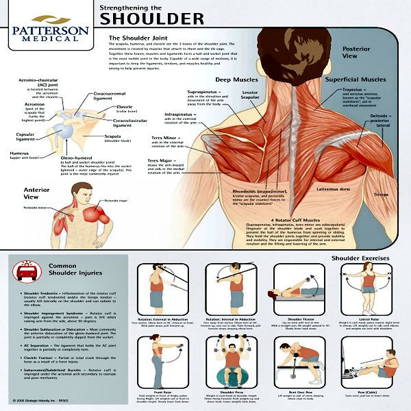
Chart Strengthening The Shoulder Joint Charts Shoulder Workout Scoliosis Exercises Workout Chart
Cpt codes for mri scans orbit face neck 70540 - wo contrast 70542 - wcontrast 70543 - wo w contrast tmj 70336 shoulder elbow or wrist upper extremity joint.
Right shoulder anatomical chart. Quantity must be between 1 and 5000. Anterior views of the muscles of the right elbow. Ad Help Educate Those In Your Institution With Our Life-Like Models Simulators. Detailed view of the socket of the right shoulder joint. The shoulder and elbow anatomy chart is 65 x 50 cm making it sufficiently large for clinic displays. One of our most popular models this functional right shoulder joint model allows demonstration of the mechanics of the shoulder and understanding of the anatomy.
Detailed view of the socket of the right shoulder joint. Right Shoulder Joint Anatomical Structure. Contact ISMA Practice Management staff at 800 257-4762 or 317 261-2060. In this image you will find clavicle collarbone bursa acromion coracoid process scapula shoulder blade in Right shoulder joint anatomical structure. Bilateral indicator of 1 must be reported with 1 unit of service and modifier 50. The shoulder is made up of two joints the acromioclavicular joint and the glenohumeral joint.
The acromioclavicular joint is where the acromion part of the shoulder blade scapula and the collar bone clavicle meet. The glenohumeral joint is where the ball humeral head and the socket the glenoid meet. Be The First To Review This Product. This angle is easily palpable and is one of the main bony land-marks at the shoulder. Illustrates posterior lateral anterior and superior views of the shoulder anatomy as well as the socket of a normal shoulder joint. The following is an overview of the shoulder muscle anatomy.
Lamination protects the surface of the chart and allows for annotation with a washable marker pen not supplied. Posterior lateral and superior views of the bones of the right shoulder. Ad Shop Preserved Specimens Specimen Displays Dissection Equipment And Dissection Guides. The acromion turns further anteriorly and covers part of the humeral head. This anatomical chart presents useful information about the shoulder and elbow. Detailed labeled illustrations of the shoulder as follows.
This Anatomy Poster shows the bones muscles ligaments veins and arteries of the shoulder. Functional Hip Joint Anatomical Model. Shoulder joint model consists of shoulder blade collar bone portion of humerus and joint ligaments. Anatomy and Injuries of the Shoulder illustrates the following normal anatomy. The shoulder joint is formed where the humerus upper arm bone fits into. Right Shoulder Joint Anatomical Structure.
33536 shoulder anatomy stock photos vectors and illustrations are available royalty-free. The creation of the pattern on the chart starts from the left shoulder. Quantity must be between 1 and 5000. Prominent and meets the acromion at a right angle posteriorly. Plus exercises for training them. You may also find long head of biceps tendon supraspinatus rotator cuff subacromial space coracoacromial ligament in it.
Ultra Realistic Medical Training Simulation Products Anatomical Models. Detailed labeled illustrations of the shoulder as follows. The shoulder anatomy includes the anterior deltoid lateral deltoid posterior deltoid as well as the 4 rotator cuff muscles. Help other Mentone Educational users shop smarter by writing reviews for products you have purchased. The tip points outwards and is easily palpated in. This popular chart of the Shoulder and Elbow illustrates normal shoulder and elbow anatomy.
It appears in a convenient poster size 50 x 67 cm 20x26 and can be written on and wiped off with non permanent markers. Posterior lateral and superior views of the bones of the right shoulder. Try these curated collections. F5 Right hand thumb F6 Right hand second digit F7 Right hand third digit F8 Right hand fourth digit F9 Right hand fifth digit TA Left foot great toe. Ad Free Shipping Available. Anatomical Chart- Shoulder and Elbow has a rating of 0 5 based on 0 reviews.
The price action then forms the head which is higher or lower depending on the type of the pattern than the left shoulder. PMD001MOD Updated 2015. The shoulder has about eight muscles that attach to the scapula humerus and clavicle. On the anterior side of the shoulder the coracobrachialis serratus anterior pectoralis major and pectoralis minor muscles work as a group to flex and adduct the scapula and humerus anteriorly toward the sternum. General bone structure and anatomy of the shoulder and elbow. Anatomical modifiers include coronary artery eye lid finger side of body and toe.
General bone structure and anatomy of the shoulder and elbow. This thickly laminated anatomical chart is printed on premium glossy 200 g UV resistant paper and comes with 2 sided lamination 75 micron. The 50 modifier identifies the service as being performed on both sides of the body. Contents hide Deltoids Anatomy. Elbow fractures icons orthopedic impingement body yoga anatomy back shoulder elbow fracture glenoid icons pain shoulder and elbow pain shoulder joint. Anterior view showing muscles bones liagments nerves veins and arterires Anterior view deep Layer of the bones ligaments and mucsle Posterior view superior and lateral views of the bones of the shoulder Detail of the right shoulder socket.
Functional FootAnkle Anatomical Models. The latissimus dorsi and teres major on the posterior side extend and adduct the arm towards the vertebrae of the back. Learn about these muscles their origin and insertion points and their functional anatomy. The muscles in the shoulder aid in a wide. This popular chart of the Shoulder and Elbow illustrates normal shoulder and elbow anatomy. See shoulder anatomy stock video clips.
The shoulder is one of the largest and most complex joints in the body. The coracoid process is found at the anterior aspect of the scapula. The right shoulder is created afterwards. Shoulder anatomy images. The Anatomy of the Shoulder. Shows impingement syndrome rotator cuff tear trauma such as proximal humeral fracture and acromio-clavicular separation and bicipital tendon problems.
These muscles form the outer shape of the shoulder and underarm.

Pin On Anatomy

Shoulder And Elbow Chart 20x26 Elbow Anatomy Joints Anatomy Anatomy

Ligaments Of The Joints Anatomical Chart Joints Anatomy Physical Therapy Human Body

Pin On Anatomy Drawings

Anatomy Lesson Shoulder Musculature Beautiful To The Core Shoulder Anatomy Muscle Anatomy Neck And Shoulder Muscles

Posterior View Of The Shoulder Shoulder Anatomy Shoulder Muscle Anatomy Muscle Anatomy

Pin On Anatomy

Joints Of The Upper Extremities Laminated Anatomical Chart Muscle Anatomy Massage Therapy Anatomy

The Muscular System Deep Layers Front Laminated Anatomy Chart Muscle Anatomy Muscular System Human Body Anatomy

Anatomy And Injuries Of The Shoulder Anatomical Chart Shoulder Anatomy Anatomy Muscle Anatomy

Anatomy Chart Muscular System Spanish Muscular System Anatomy Muscular System Muscle Anatomy

Anatomical Charts And Posters Anatomy Charts Arm And Leg Charts Shoulder And Elbow Laminated Chart Shoulder Anatomy Anatomy Joints Anatomy

Taking It To The Next Level With The Dailey Method Shoulder Anatomy Muscle Diagram Shoulder Muscles

Shoulder And Elbow Chart 20x26 Elbow Anatomy Joints Anatomy Anatomical
Posting Komentar untuk "Right Shoulder Anatomical Chart"