Will Mri Show Torn Muscle
This may limit peoples ability to brush their hair or put on clothing. Gumina et al on 586 patients with a history of arthroscopic tear repair reported a mean age of 59 yearsPatients older than 60 were twice as likely to develop tear which were larger and more massiveThe prevalence in human.

Shoulder Anatomy Mri Joints Anatomy
The peroneus longus muscle originates from the posterolateral condyle of the tibia the interosseous membrane and the proximal fibula.

Will mri show torn muscle. Advancing age has been consistently held accountable as one of the major risk factor for the development of cuff tears in various studies. Aetna considers magnetic resonance imaging MRI studies of the knee medically necessary when any of the following criteria is met. On axial MRI a partially torn subluxed tendon is seen medial or anterior to the medial malleolus. Magnetic resonance imaging MRI. In type 2 torn pectoral the tear occurred where the muscle starts transitioning into a tendon the muscle-tendon junction- harder to fix but still possible. And while muscle does show up on some scans it will only show a problem if there is a tear or A LOT of inflammation.
Symptoms may include shoulder pain which is often worse with movement or weakness. However magnetic resonance imaging Computed tomography CT and magnetic resonance imaging MRI A doctor can often diagnose a musculoskeletal disorder based on the history and the results of a physical examination. MRIs are used for men to determine prostate cancer and guide biopsies of the prostate. In type 1 torn pectoral the tendon of the pectoralis major tears directly off of the humerus the easiest to fix. Calf muscle tears usually heal after a few weeks of conservative treatments such as rest ice compression and elevation. Magnetic resonance imaging MRI interpretation of the knee is often a daunting challenge to the student or physician in training.
He or she inserts a tool to grasp the end of the latissimus. The glenohumeral joint is a synovial joint formed by the glenoid fossa of the scapula and the head of the humerus while the acromioclavicular joint connects the acromion and the. A rotator cuff tear is an injury where one or more of the tendons or muscles of the rotator cuff of the shoulder get torn. In cases of tendinitissuch as that seen with jumpers kneean MRI will usually reveal progressive knee injury in the form of scarring inflammation and malformations of the tendon. The harder or more calcified something is the better it shows up on x-rays therefore x-rays are very. Laboratory tests imaging tests or other.
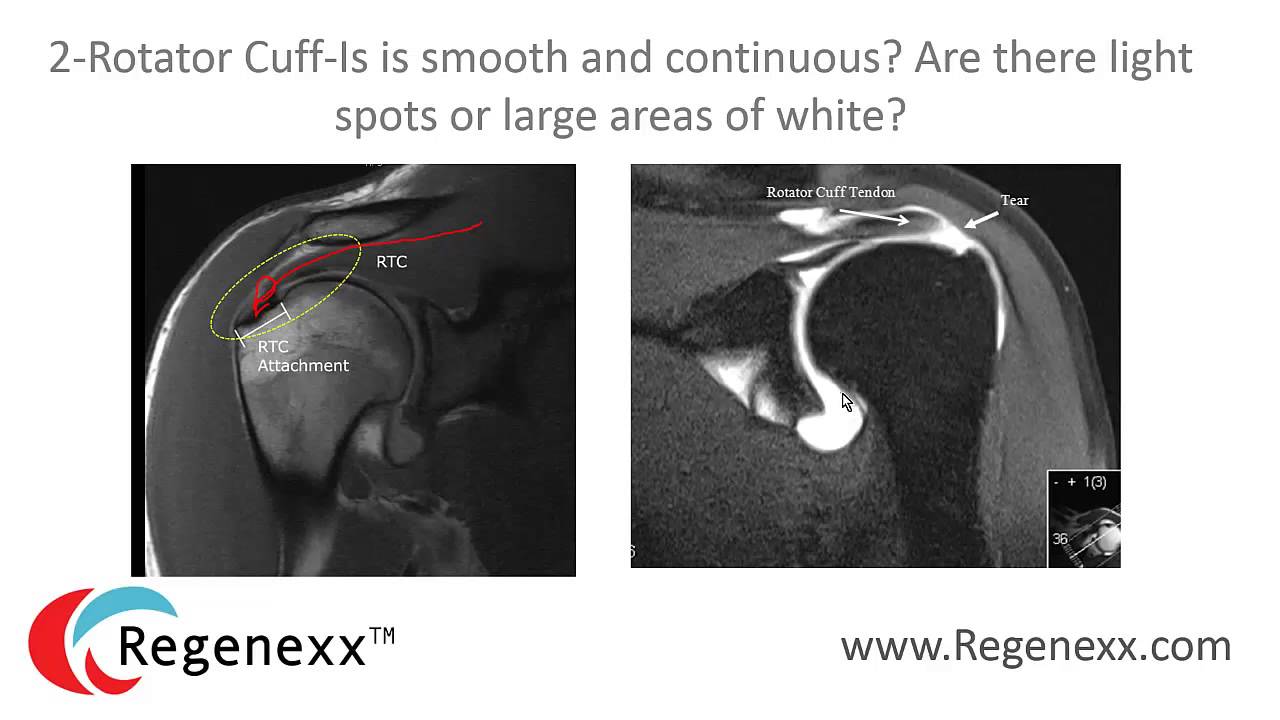
Normal shoulder MRI. In other words the scan is like cutting cheese in that they do not see the. An MRI scan uses a strong magnetic field and radio waves to create pictures on a computer screen. Magnetic Resonance Imaging MRI- An MRI can invasively way to show any hairline fracture or tears in the rib cage. On images of the shoulder with the arm in a neutral position the torn labrum may be held in its normal anatomic position by the intact scapular periosteum which thereby prevents contrast media from entering the tear. Soft tissue parts like muscle and skin show up as a gray shadow on plain x-ray.
While MRIs can help your doctor. MRI or diagnostic ultrasound may be needed to best diagnose the location and degree of the injury. An MRI scan is a detailed way of looking at the inside of the body. Athletes and people over 40 are especially prone to this type of muscle injury. Ravens cornerback Marlon Humphrey a team leader and one of the NFLs top defensive backs will miss the rest of the season after tearing a pectoral muscle in Sundays loss to the Pittsburgh. For most people that have pain it is caused by muscle imbalances not anything that can be surgically repaired or can be seen on imaging.
ACL surgery is a repair or reconstruction of the anterior cruciate ligament ACL. Most problems that cause every day aches and pains are muscle related. Bone Scan- This test is more in-depth than an x-ray and allows a closer look at any damage of the rib. Clicking may also occur with movement of the arm. MRI which uses radio waves and a powerful magnet to make detailed pictures of your shoulder. Tear 2 the number of tendons torn 3 if it is the dominant or non-dominant arm involved 4.
It can show tissues organs and other structures inside your body. In type 3 pec ruptures the tear is completely within the muscle. X-Ray- A chest x-ray can help rule out any extensive damage to the ribs but it will not pick up small tears or fractures. A partially or completely torn ACL is a common injury among athletes. The ACL is an important soft-tissue structure in the knee that connects the femur to the tibia. 1-4 It is well known that the atrophy of rotator cuff muscles is one.
Arthrography can be used to view torn ligaments and fragmented cartilage in the joint. Grade II strains involve more extensive damage up to 90 of the muscle fibers may be torn. A torn calf muscle is a painful injury in the muscles behind your shin bone. The articularis genus muscle the final component of extensor mechanism arises from the distal portion of the femur and inserts into the suprapatellar pouch. Or Persistent knee painswelling andor instability giving way not associated with an injury and not responding to at least 3 weeks of. The postoperative chronically torn ACL graft may.
Muscle atrophy means a gradual shrinking of the muscle tissue as a result of inactivity or disease and atrophy of the rotator cuff muscle may occur for a variety of reasons such cuffas aging disuse diabetes and suprascapular nerve injury with the most common being chronic rotator cuff tears. Muscle Deltoid muscle. The persons eyeballs can be seen at the top of the picture. These injuries present with more pain described as sharp in nature significant loss of muscle strength and range of motion. X-rays to see if the top of your arm bone humeral head is pushing into your rotator cuff space. You wont see it very well on an x-ray.
It is composed of two articulations. In the front the surgeon creates a flap in the deltoid muscle which covers the shoulder. Detection staging and post-treatment evaluation of tumor of the knee. This image is an MRI scan of a brain. Abdominal MRIs can look for cancerous tumors and show certain liver spleen kidney and pancreas diseases. An MRI will be able to take pictures of cartilage and ligaments to determine if theres a meniscus tear.
The glenohumeral and acromioclavicular joints. MRI stands for magnetic resonance imaging. A tendon is the fibrous tissue that attaches muscle to bone in the human body. The shoulder joint is a joint that connects the upper limb to the axial skeleton. An MRI can be used to detect chronic tendinitis inflammation of the tendon or tendon ruptures although this is usually apparent on physical examination. The forces applied to a tendon may be more than 5 times your body weight.
Although a rotator cuff tear wont show up on an X-ray this test can visualize bone spurs or other potential causes for your pain such as arthritis. An MRI of a joint can show torn ligaments muscle tears or arthritis. MRI stands for magnetic resonance imaging. The radiologist gets the scan on the computer and the MRI images show them different parts of the shoulder in slices. This means that MR-arthrography with the arm in the neutral position may fail to detect the labral tear.

Maurkice Pouncey Torn Mcl Mri Sports Injury Mcl Anatomy Of The Knee

Pin On Back Pain Alternative Treatment Options

Pin By Eenee Dct On Bone And Joints Knee Mri Medical Terms Bone And Joint
Pin On Labrum And Rotator Cuff Tear Pt

Pin On Mri

Pin On Randy Shank Rolfing

Pin By Dr Abuaiad On Bone And Joints Mri Brain Knee Mri Bone And Joint

Pin By Dr Abuaiad On Bone And Joints Radiology Imaging Anatomy Of The Knee Knee Mri
Biceps Tendon At 12 O Clock And Does Not Extend To The 1 3 O Lock Position A Shoulder Anatomy Mri Shoulder Joint Anatomy

Pin On Mri

Pin On Labrum And Rotator Cuff Tear Pt

Lumbar Spine Image Medical Anatomy Radiology Imaging Medical Knowledge

Shoulder Rotator Cuff Tear Mri Unidad Especializada En Ortopedia Y Traumatologia En Bogota Colombia Pbx 6923370 Www U Mri Shoulder Anatomy Radiology Imaging

How Do I Diagnose A Torn Rotator Cuff One Way Is Through An Mri Magnetic Resonance Imaging If An Rotator Cuff Tear Rotator Cuff Torn Rotator Cuff Symptoms
Posting Komentar untuk "Will Mri Show Torn Muscle"