Diagnostic Ultrasound For Neck Pain
Clinical Radiology Vol75 No5 p341-347. Part 2 Upper Limb on Itunes A Practical Guide to Ultrasound Imaging For Regional Anesthesia.

Stretches For Sonographers Sonography Ultrasound Tech Sonographer
Medical ultrasound includes diagnostic techniques mainly imaging techniques using ultrasound as well as therapeutic applications of ultrasound.
Diagnostic ultrasound for neck pain. In diagnosis it is used to create an image of internal body structures such as tendons muscles joints blood vessels and internal organs to measure some characteristics eg. Many cases of neck pain are diagnosed based on the patients medical history and physical exam. AMA PRA Category 1 CME credit for Clinical Imaging reviewers. Some ultrasound examinations require special preparation beforehand such as. Shoulder pain evaluate for rotator cuff abnormality Findings. It is used to help diagnose sprains strains tears trapped nerves arthritis and other musculoskeletal conditions.
Ultrasound imaging or magnetic resonance neurography. Diagnostic testing would be recommended for anyone at risk for a stroke to check for blockages in the carotid artery said McLeod Vascular Surgeon Dr. Ultrasound is safe noninvasive and does not use ionizing radiation. Her diagnostic skills and her rapport with patients separates her from doctors I have seen in the past. Image guidance with fluoroscopy or ultrasound should be used for cervical medial branch blocks grade A recommendation moderate level of certainty. Pregnancy ultrasound is used to look at an unborn baby.
Neck masses that are located on the anterior neck should have ultrasound Procedure 76536 performed as the initial imaging study. Chazz Michael Michaels Registration Number. CT scans of the head and neck may show silent lacunar strokes undiagnosed strokes creating cavities after the stroke has healed doppler ultrasound of the neck may identify stenosis caused by lesions as well as. Ultrasound Exams Diagnostic codes. The test can provide information about a babys growth development and. A special type of ultrasound scan called a Doppler ultrasound is used to detect the speed and direction of blood flow in certain regions of the body for example neck arteries and leg veins.
This tensile stress is supported by the medium and for example a 2-MPa rarefactional pressure which is common even for diagnostic ultrasound represents a negative tension 20 times atmospheric pressure i. The images can provide valuable information for diagnosing and treating a variety of diseases and conditions. The specialist might also look at the necks blood flow using an X-ray CT or MRI image angiogram. Tima Le has treated me over the years for various conditions associated with my neck back and spine. A Practical Guide to Ultrasound Imaging For Regional Anesthesia. Neck to Shoulder pain can be caused by a number of factors including muscle strain ligament sprains arthritis or a pinched nerve.
Imaging manifestations and diagnostic value of chest CT of coronavirus disease 2019 COVID-19 in the Xiaogan area Wang et al. When lumbar or leg pain increase during the SLR test with dorsiflexion of the ankle or flexion of the neck a neural pain source is alleged to be. There are two main categories of ultrasounds. For possible neck masses or fullness of the neck that is not well described on physical examination ultrasound Procedure 76536 or ENT evaluation can be helpful in making decisions regarding the need for advanced imaging. Ultrasound transmitted into a tissue may have rarefactional pressure amplitudes of several megaPascals MPa. There is a focal anechoic tear of the anterior distal aspect of the supraspinatus tendon measuring 1 cm short axis by 15 cm long axis.
Youll receive a local anesthetic to numb the area on your neck. Medical issues and ultrasound scans. And ultrasound US can be used to identify nerve derangement and rupture and neuroma formation. The ultrasound image shows the size of the prostate and any abnormal-looking areas such as tumors. An ultrasound can also show parts of the body in motion such as a heart beating or blood flowing through blood vessels. There is significant room for improvement in the development of more formal diagnostic tools aiding prognostication for these difficult and sometimes severe injuries.
Ultrasound of the Shoulder Date of Study. To determine whether a tumor is cancerous the health care provider uses the transducer and ultrasound images to guide a needle to the tumor. In those with chronic neck pain conservative treatment should be used prior to prognostic cervical facet blocks grade B recommendation moderate level of certainty. 6821 Other cellulites and abscess of neck 7842 Swelling mass or lump in head and neck. An Illustrated and Clinically Orientated Guide. Best-evidence diagnostic rules based on systematic reviews.
March 11 2017 Patient Name. Considers non-operational adult ultrasound of the spine and paraspinal tissues for the evaluation of spine and paraspinal tissues for the evaluation of neuromuscular conditions and all other indications for example to assist in lumbar puncture or to assist with interventional pain injections non-covered CPT Diagnostic Ultrasound. Ultrasound Exam CPT code s Abdominal complete 76700. Diagnostic ultrasound also called sonography or diagnostic medical sonography is an imaging method that uses high-frequency sound waves to produce images of structures within your body. Various imaging technologies are available to give a better view of what might be causing neck pain. When more information is need advanced diagnostics may be needed to reach an accurate diagnosis.
Neck Pain and Yawning Diagnosis. Highland EM Ultrasound Fueled pain management Common ED Injury clavicle fracture Shoulder dislocation Humeral Fracture distal radius fracture Boxers fracture palmar hand laceration Anterior Lateral Rib Fracture Posterior Rib Fracture Hip FxFemur Fx pubic rami fracture distal tibiafib fracture bimall fracture sole of foot laceration. Le is extremely thorough and takes the time to listen to her patients. The Editors of Clinical Imaging in conjunction with the Elsevier Office of Continuing Medical Education are pleased to offer an AMA PRA Category 1 CME credit program for registered Clinical Imaging physician reviewers who complete manuscript reviews. During the procedure the doctor will use ultrasound to view your jugular vein and will use fluoroscopy a type of x-ray imaging to view the veins between your neck and your liver. 78650 Chest pain unspecified 78651 Precordial pain 79431 Abnormal EKG.
In addition cost. Transrectal ultrasound cannot definitively identify prostate cancer. Distances and velocities or to generate an. Part 3 Lower Limb on Itunes Ultrasound for Pain Medicine Intervention. Approximately 10 percent of adults have neck to Shoulder pain at any one time. Image guidance with fluoroscopy or ultrasound should be used for cervical medial branch blocks grade A recommendation moderate level of certainty.
The EOCME is accredited by the Accreditation Council for. Clinical classification in low back pain. A variety of diagnostic tests and scans may be used to identify the nature and source of neck pain and other symptoms of yawning. Tom Petersen 1 Mark Laslett 2. Ultrasound imaging uses sound waves to produce pictures of muscles tendons ligaments nerves and joints throughout the body. Pain Neuromuscular Medicine Rehabilitation.
In those with chronic neck pain conservative treatment should be used prior to prognostic cervical facet blocks grade B recommendation moderate level of certainty. Pregnancy ultrasound and diagnostic ultrasound. The majority of patients regardless of the cause of pain recover with conservative Physiotherapy treatment. A Practical Guide Basic Science of Pain.

Pin On Medical

Simtics Ultrasound Technician Diagnostic Medical Sonography Sonography

Pedunculated Fibroid Diagnostic Medical Sonography Medical Ultrasound Fibroids

Neck Ultrasound Ultrasound Ultrasound Technician Sonography

Pin On Ultrasound Notes

Mirizzi Syndrome Radiology Case Radiopaedia Org Diagnostic Medical Sonography Radiology Ultrasound

Pin On Physical Therapy

Pin By Dr Abuaiad On Lymphatics Diagnostic Medical Sonography Nuclear Medicine Sonography
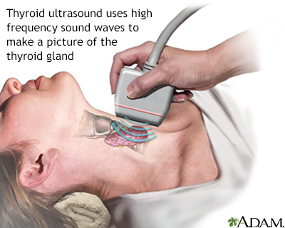
Thyroid Ultrasound Information Mount Sinai New York

What Is A Neck Ultrasound Two Views

Pin On Superficial

Differential Diagnosis Of A Cystic Pediatric Neck Mass Learn It Here Http Radiologypics Com 2013 03 06 Different Pediatrics Diagnosis Ultrasound Sonography

Neck Ultrasound Sjogren Syndrome Multiple Hypoechoic Fossi Due To Lymphocytic Infiltrations Diagnostic Medical Sonography Ultrasound Stroke Prevention

Pin By Dr Abuaiad On Superficial Medical Ultrasound Ultrasound Sonography Ultrasound
Posting Komentar untuk "Diagnostic Ultrasound For Neck Pain"