Eeg Vs Mri For Seizures
Tonic seizures manifest with sustained contraction of facial limb axial and other muscles. Concurrent risk factors including preexisting epilepsy structural brain lesions and the use of drugs contribute to the development of seizures in many patients with AWS.

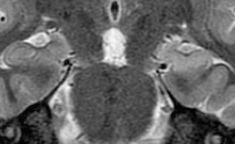
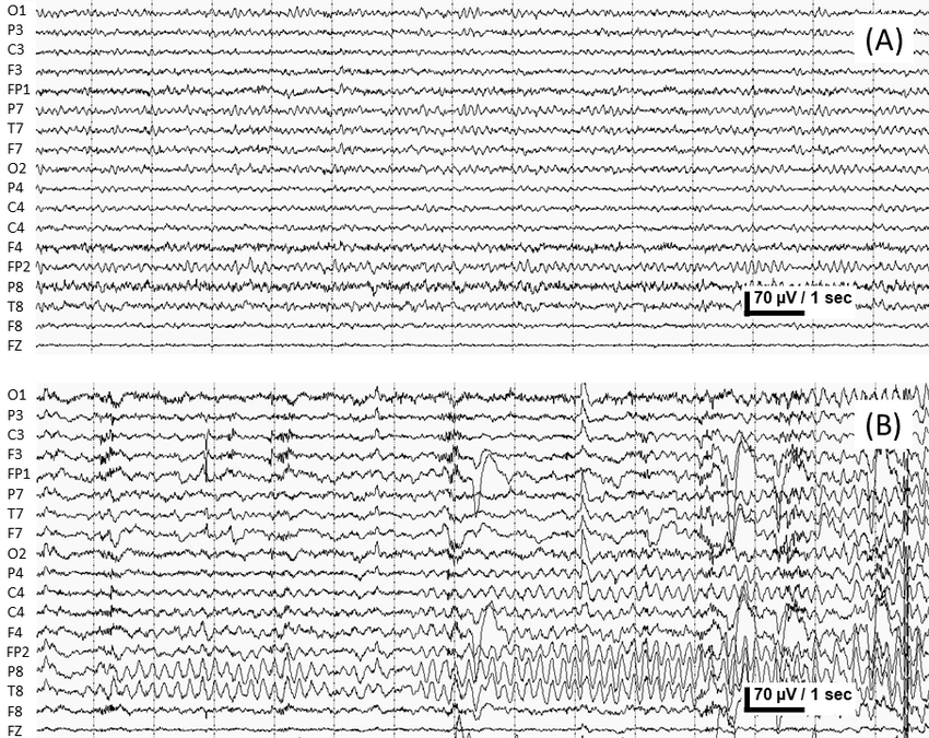
The Eeg Mri And Seizure Frequency For Patient 2 A Interictal Eeg Download Scientific Diagram
In the electrographic group EEG-detected seizures fulfilling diagnostic criteria were treated if they lasted more than 2 minutes or occurred more than twice in 24 hours.

Eeg vs mri for seizures. Absence seizures most commonly affect children between the ages of 4 and 12They can also occur in adults. 30516599 For patients with SAH near the base of the brain and no defined source of bleeding cervical spine MRI may be useful to evaluate for a spinal source of bleeding. Other methods to study brain function include. A standard EEG can detect seizures and diagnose epilepsy but a sleep-deprived EEG may better detect more subtle seizures like absence seizures or focal seizures. Clonic seizures are rhythmic jerks that may localise in a small part of the face or limbs axial muscles and the diaphragm or be multifocal or hemiconvulsive. An electroencephalogram EEG is a non-invasive test that records electrical activity in the brain.
The incidences of focal seizures focal neurologic deficits and focal imaging abnormalities are lower. Electroencephalography EEG is a simple and painless study that records the brains electrical activity picked up by tiny wires taped to the head. Uses a portable EEG machine and sophisticated built-in algorithms identifies EEG patterns within 24 hours of injury that are associated with structural brain injury visible on CT scans. Neurologists can use EEG results to identify abnormal electrical activity in the brain and diagnose certain conditions such as. MRI can be useful to detect subtle underlying pathology eg arteriovenous malformations infections malignancy or inflammatory disorders. Patients and caregivers are naturally worried when.
MRI is more sensitive than CT and is therefore preferred especially for the detection of cortical malformation dysgenesis or hippocampal sclerosis. Functional magnetic resonance imaging fMRI. The journals editor Yasmin Khakoo MD FAAN in conjunction with the. Other Brain Measurement Techniques. Imaging tests including CT scans MRI scans and EEG scans are sometimes used to check for abnormalities in the nasal cavity brain or nervous system. In general MRI is the preferred technique because it.
Epilepsy is the medical name given to the condition in which you experience recurrent seizures. The advantage of EEG measurement is that it is the least invasive measure of brain activity we have available and provides lots of quantitative information during relevant cognitive processes. The primary outcome was. In this series the clinical correlates of BiPLEDs differed somewhat from those of PLEDs. The journal has a broad International perspective and emphasises the advances occurring in Asia the Pacific Rim region Europe. One of the 9 patients had HIV coinfection.
Defining Seizures and Epilepsy. The MRI findings in 2 of the 9 patients showed edema. This physician will also conduct a thorough neurological examination. Continuous EEG electroencephalogram monitoring may be needed to monitor the seizures and how a person responds to treatment. EEG is not indicated if patients have. The timing of seizures in relation to triggers or the presence of a discernible trigger is the basis for their classification as provoked or unprovoked.
This International journal Journal of Clinical Neuroscience publishes articles on clinical neurosurgery and neurology and the related neurosciences such as neuro-pathology neuro-radiology neuro-ophthalmology and neuro-physiology. MRI white matter lesions Many times I get consulted by patients or their relatives when their MRI brain report reads multiple scattered white matter lesions seen. At times medicines called anesthetics are used in the hospital to put a person into a coma to stop the seizures. The incidence of coma is higher in BiPLEDs than in PLEDs 72 vs 24 and the mortality rate is higher 61 vs 29. EEGs are usually done to detect seizures and to diagnose epilepsy but they can be used to evaluate or diagnose other conditions such as sleep disorders or brain injuries. MRI is more likely to show an abnormality in a patient with focal seizures abnormal neurologic findings or focal discharges on EEG.
Pediatric Neurology publishes timely peer-reviewed clinical and research articles covering all aspects of the developing nervous systemPediatric Neurology features up-to-the-minute publication of the latest advances in the diagnosis management and treatment of pediatric neurologic disorders. Wasay et al using EEG and MRI to study patients found that of the 9 patients who were examined with EEG all 9 had seizures or other abnormalities and 1 had nonconvulsive status epilepticus. Todds paresis follows prolonged hemiconvulsions. Magnetic resonance imaging MRI and computed tomography CT of the head can be used to evaluate patients with seizures. Absence seizures tend to cause shorter and milder symptoms than tonic-clonic seizures. They may be focal multifocal or generalised symmetrical or.
Seizures Decorticate vs Decerebrate posturing Decerebrate posturing is an abnormal body posture and it is defined the arms and legs being held straight out the toes being pointed downward and the head and neck being arched backward 3. 20 EEG is recommended in new-onset seizures or when showing a new pattern in patients with a known history of alcohol-related seizures. Provoked seizures are those that occur within 7 days of an acute neurologic systemic metabolic or toxic eg visual intoxication or detoxication insult. Learn about sleep-deprived EEGs their purpose in diagnosing seizures potential risks and costs and what to expect before during and after the testing is completed. Specific brain wave patterns may be noted during or between seizures in patients with epilepsy and may help with diagnosis. In most cases an EEG electroencephalogram and MRI magnetic resonance imaging test will be performed as well.
The radiologists report usually further reads that these can be seen in primary demyelinating conditions like multiple sclerosis or in vascular disorders. When these seizures are tied to another event like drug or alcohol withdrawal the underlying. This is interesting as beta activity is a rare ictal EEG pattern observed sometimes in specific subgroups such as patients suffering from seizures due to A-V malformations porencephalic cysts or tumors in children with LennoxGastaut Syndrome or infantile spasms ie showing little overlap with our sample. Tests may also be needed to find the cause of the seizure emergency so it can be treated correctly. It works by picking up abnormal brain waves via electrodes that are attached to the scalp. The first step is to review your medical history including a detailed recounting of the seizures with your physician.

Diagnosis Epilepsy Action Australiaepilepsy Action Australia
:max_bytes(150000):strip_icc()/sleep-deprived-eeg-and-seizures-4628312_final-a8f1e5e503d04324918753e48f6d1521.png)
How A Sleep Deprived Eeg May Diagnose Seizures

Eeg In Epilepsy Medlink Neurology

The Merits Of Meg In Children A Noninvasive Means To Refine Epilepsy Localization Consult Qd

Eeg Fmri In A Patient With Left Temporal Lobe Epilepsy And Left Download Scientific Diagram

Mri And Eeg Could Identify Children At Risk For Epilepsy After Febrile Seizures National Institutes Of Health Nih
Which Imaging Exam Is Ideal For The Diagnosis And Treatment Of Epilepsy Ucsf Radiology
Pin On Urination Or Micturition Epilepsy

Normal Eeg Awake Compared To Lennox Gastaut Syndrome Epilepsy Awareness Month Epilepsy Awareness Syndrome
Normal Eeg Compared To Eeg Including A Seizure A Normal Eeg Of 15 Download Scientific Diagram

Photographic Print Epilepsy Mri Brain Scan And Eeg Trace By Pasieka 24x18in In 2021 Brain Scan Mri Brain Brain Art

Eeg Vs Mri Vs Fmri What Are The Differences

Infographic First Seizure Management Infographic Epilepsy Types Seizures

Source Imaging Of Seizure Onset Predicts Surgical Outcome In Pediatric Epilepsy Sciencedirect
Posting Komentar untuk "Eeg Vs Mri For Seizures"