Posterior Fossa Syndrome Prognosis
Whole tendon disinsertion of superior oblique for the treatment of Brown syndrome. Posterior circulation ischaemic stroke is a clinical syndrome associated with ischaemia related to stenosis in situ thrombosis or embolic occlusion of the posterior circulation arteriesthe vertebral arteries in the neck the intracranial vertebral basilar and posterior cerebral arteries and their branches fig 1.

Posterior Fossa Syndrome Review Of The Behavioral And Emotional Aspects In Pediatric Cancer Patients Lanier 2017 Cancer Wiley Online Library
The sphenoid bone is an unpaired bone of the neurocraniumIt is situated in the middle of the skull towards the front in front of the basilar part of the occipital boneThe sphenoid bone is one of the seven bones that articulate to form the orbitIts shape somewhat resembles that of a butterfly or bat with its wings extended.

Posterior fossa syndrome prognosis. A comparative study with multiple techniques. Bordarier C Aicardi J. Pediatric Neurology publishes timely peer-reviewed clinical and research articles covering all aspects of the developing nervous systemPediatric Neurology features up-to-the-minute publication of the latest advances in the diagnosis management and treatment of pediatric neurologic disorders. Arachnoid cysts can be primary or secondary. 101210jc2013-3487 28054130 Fischer M Schmutzhard E. The mission of The Annals of Thoracic Surgery is to promote scholarship in cardiothoracic surgery patient care clinical practice research education and policy.
Reversible posterior leukoencephalopathy syndrome RPLS is a clinical radiographic syndrome of heterogeneous etiologies that are grouped together because of similar findings on neuroimaging studies. The lens is considered subluxed when it is partially displaced but remains within the lens space. Dandy-Walker syndrome and agenesis of the cerebellar vermisdiagnostic problems and genetic counseling. Cerebellar infarctions can rapidly swell due to cytotoxic edema within the tight constraints of the non-yielding posterior fossa and lead to obstructive hydrocephalus or fatal herniation if not adequately managed in a timely fashion 117119 Figures Figures4DF. This International journal Journal of Clinical Neuroscience publishes articles on clinical neurosurgery and neurology and the related neurosciences such as neuro-pathology neuro-radiology neuro-ophthalmology and neuro-physiology. As the official journal of two of the largest American associations in its specialty this leading monthly enjoys outstanding editorial leadership and maintains rigorous selection standards.
This monthly journal offers comprehensive coverage of new techniques important developments and innovative ideas in oral and maxillofacial surgeryPractice-applicable articles help develop the methods used to handle dentoalveolar surgery facial injuries and deformities TMJ disorders oral cancer jaw reconstruction anesthesia and analgesiaThe journal also. Post stroke considerations for the posterior fossa. Posterior reversible encephalopathy syndrome PRES Reversible posterior cerebral edema syndrome. Cerebellar edema with clinical deterioration is generally regarded as an indication for suboccipital crainectomy. Primary arachnoid cysts are congenital present at birth resulting from abnormal development of the brain and. Lateral medullary syndrome LMS also called Wallenberg syndrome or posterior inferior cerebellar artery syndrome results from a vascular event in the lateral part of the medulla oblongata.
Dev Med Child Neurol 1990324. The lens is defined as luxated dislocated when it lies completely outside of the hyaloid fossa is free-floating in the vitreous is in the anterior chamber or lies directly on the retina. Dichoptic visual field mapping of suppression in exotropia with homonymous hemianopia. Ectopia lentis is the dislocation or displacement of the natural crystalline lens. Cubital tunnel syndrome is a progressive entrapment neuropathy of the ulnar nerve at the medial aspect of the elbow. I prefer the middle cranial fossa approach for several reasons.
DandyWalker malformation DWM also known as DandyWalker syndrome DWS is a rare congenital brain malformation in which the part joining the two hemispheres of the cerebellum the cerebellar vermis does not fully form and the fourth ventricle and space behind the cerebellum the posterior fossa are enlarged with cerebrospinal fluidMost of those affected. J Clin Endocrinol Metab. You have to assume that you are operating on either side of it when you drill open the semicircular canal and plug it. The American Journal of Ophthalmology is a peer-reviewed scientific publication that welcomes the submission of original previously unpublished manuscripts directed to ophthalmologists and visual science specialists describing clinical investigations clinical observations and clinically relevant laboratory investigations. The ulnar nerve which is a motor and sensory nerve is formed from the medial cord of the brachial plexus which originates from nerve roots C8 and T1. He first reported the case of LMS.
Dandy-Walker complex is a group of disorders that affect the development of the brainThe changes in brain development are present from birth congenitalDandy-Walker complex affects the formation of the area of the brain known as the cerebellum which is responsible for coordinating movement and the fluid-filled spaces around itPeople with Dandy. The strict definition of the Ramsay Hunt syndrome is peripheral facial nerve palsy accompanied by an erythematous vesicular rash on the ear zoster oticus or in the mouth. Cerebellar infarction is problematic due to the low volume of the posterior fossa. Our doctors define difficult medical language in easy-to-understand explanations of. J Ramsay Hunt who described various clinical presentations of facial paralysis and rash also recognised other frequent symptoms and signs such as tinnitus hearing loss nausea vomiting vertigo and. First you can see the dehiscence directly with the middle cranial fossa approach.
Arachnoid cysts are sacs filled with cerebrospinal fluid CSF that are located between the brain or spinal cord and the arachnoid membrane one of the three membranes that cover the brain and spinal cord. It was named after Adolf Wallenberg 1862-1949 who was a renowned Jewish neurologist and neuroanatomist who practiced in Germany. Supraspinatus tendinosis refers to the intratendinous degeneration of the supraspinatus tendon that is thought to be a result of chronic overuse and that does not have a significant inflammatory component 1Supraspinatus tendinosis and tendon tears is mostly between the fifth to sixth decades of life with the size of the tear increasing with age 2. Posterior reversible encephalopathy syndrome due to malignant hypercalcemia. The ulnar nerve travels down the posterior aspect of the arm to eventually traverse posterior to the medial. It is also often referred to as.
The journals editor Yasmin Khakoo MD FAAN in conjunction. Swelling may compress the brainstem or the cerebral aqueduct causing noncommunicating hydrocephalus. The journal has a broad International perspective and emphasises the advances occurring in Asia the Pacific Rim region Europe. There are four categories of thoracic outlet syndrome and each presents with unique signs and symptoms see Table 1. Barkovich A Kjos B Norman D et al. Revised classification of posterior fossa cysts and cystlike malformations based on the results of multiplanar MR imaging.
A CT scan demonstrated a 10 x 65 x 73 cm pelvic mass arising in the vicinity of the prostate inseparable from the posterior wall of the bladder and anterior wall of the rectum obstructing the right ureter and causing right hydronephrosis with associated bilateral external and left internal iliac adenopathy. With the transmastoid approach you cannot see the dehiscence. Patients with lower plexus C8 T1 involvement typically have symptoms that are present in the anterior and posterior shoulder region and radiate down the ulnar side of the forearm into the hand the ring and small fingers.

Prenatal Imaging Of Posterior Fossa Disorders A Review European Journal Of Paediatric Neurology
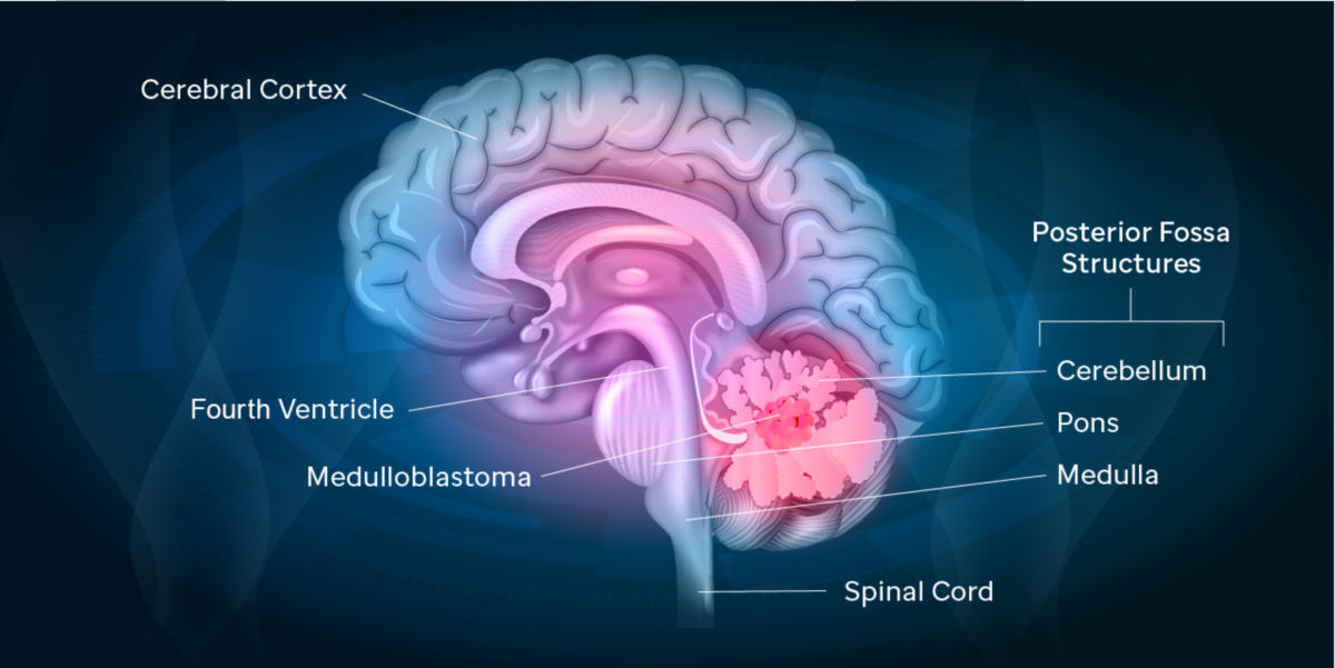
Brain Stem And Posterior Fossa John R Hesselink Md Facr And John F Healy Md Facr The Posterior Fossa Houses The Brainstem And Cerebellum The Brainstem Contains All The Cranial Nerve Nuclei And Many Efferent And Afferent Fiber Tracts

Posterior Fossa Syndrome Review Of The Behavioral And Emotional Aspects In Pediatric Cancer Patients Lanier 2017 Cancer Wiley Online Library
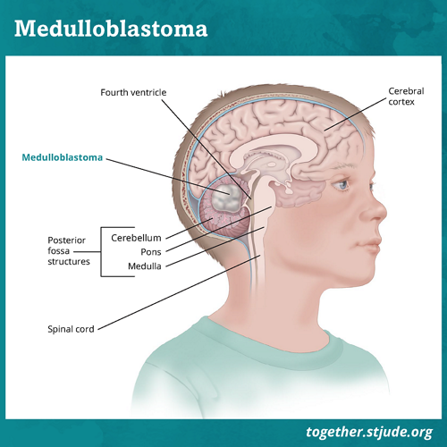
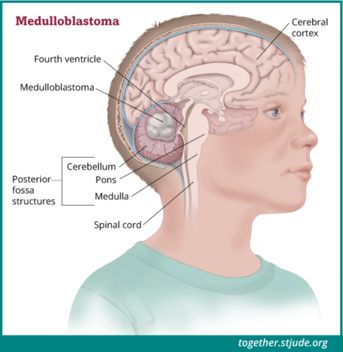
Posterior Fossa Syndrome Together

Posterior Fossa Syndrome Together

Posterior Fossa Tumors In Children Radiological Tips Tricks In The Age Of Genomic Tumor Classification And Advance Mr Technology Sciencedirect
Posterior Fossa Syndrome Together

The Mcpprh Score In Children With Posterior Fossa Neoplasms Download Table

Flow Chart Of A General Algorithm For Management Of Posterior Fossa Download Scientific Diagram
Dana Farber Org

Posterior Fossa Malformations Neuroimaging Clinics
Getting To The Bottom Of A Medulloblastoma Mystery St Jude Progress
The Other C Word Child Aya Cancer Awareness Life Interrupted Child Cancer Awareness 2020 Brain Surgery And Posterior Fossa Syndrome Posterior Fossa Syndrome Involves A Variety Of Signs And


Schematic Illustration Of The Relationship Between The Posterior Fossa Download Scientific Diagram
Posting Komentar untuk "Posterior Fossa Syndrome Prognosis"